Explore
 Validate
 Learn
Learn Western blot
 Immunoprecipitation
Antibody data
- Antibody Data
- Antigen structure
- References [10]
- Comments [0]
- Validations
- Western blot [1]
- Flow cytometry [5]
- Other assay [4]
Submit
Validation data
Reference
Comment
Report error
- Product number
- MA5-11895 - Provider product page

- Provider
- Invitrogen Antibodies
- Product name
- CD47 Monoclonal Antibody (B6H12.2)
- Antibody type
- Monoclonal
- Antigen
- Purifed from natural sources
- Description
- MA5-11895 targets CD47 in WB, FACS, IF, and IP applications and shows reactivity with Human samples. The MA5-11895 immunogen is intact CD47 purified from placenta.
- Reactivity
- Human
- Host
- Mouse
- Isotype
- IgG
- Antibody clone number
- B6H12.2
- Vial size
- 500 μL
- Concentration
- 0.2 mg/mL
- Storage
- 4°C
Submitted references Oxidation of Innate Immune Checkpoint CD47 on Cancer Cells with Non-Thermal Plasma.
The ionophore antibiotic gramicidin A inhibits pancreatic cancer stem cells associated with CD47 down-regulation.
Leishmania donovani Induces Autophagy in Human Blood-Derived Neutrophils.
The "don't eat me" signal CD47 is a novel diagnostic biomarker and potential therapeutic target for diffuse malignant mesothelioma.
A cytokine-controlled mechanism for integrated regulation of T-lymphocyte motility, adhesion and activation.
Latency associated peptide has in vitro and in vivo immune effects independent of TGF-beta1.
Increasing survival of ischemic tissue by targeting CD47.
Erythrocyte adhesion is modified by alterations in cellular tonicity and volume.
The C-terminal 26-residue peptide of serpin A1 stimulates proliferation of breast and liver cancer cells: role of protein kinase C and CD47.
Increased erythrocyte adhesion in mice and humans with hereditary spherocytosis and hereditary elliptocytosis.
Lin A, Razzokov J, Verswyvel H, Privat-Maldonado A, De Backer J, Yusupov M, Cardenas De La Hoz E, Ponsaerts P, Smits E, Bogaerts A
Cancers 2021 Feb 2;13(3)
Cancers 2021 Feb 2;13(3)
The ionophore antibiotic gramicidin A inhibits pancreatic cancer stem cells associated with CD47 down-regulation.
Wang RQ, Geng J, Sheng WJ, Liu XJ, Jiang M, Zhen YS
Cancer cell international 2019;19:145
Cancer cell international 2019;19:145
Leishmania donovani Induces Autophagy in Human Blood-Derived Neutrophils.
Pitale DM, Gendalur NS, Descoteaux A, Shaha C
Journal of immunology (Baltimore, Md. : 1950) 2019 Feb 15;202(4):1163-1175
Journal of immunology (Baltimore, Md. : 1950) 2019 Feb 15;202(4):1163-1175
The "don't eat me" signal CD47 is a novel diagnostic biomarker and potential therapeutic target for diffuse malignant mesothelioma.
Schürch CM, Forster S, Brühl F, Yang SH, Felley-Bosco E, Hewer E
Oncoimmunology 2017;7(1):e1373235
Oncoimmunology 2017;7(1):e1373235
A cytokine-controlled mechanism for integrated regulation of T-lymphocyte motility, adhesion and activation.
Bergström SE, Bergdahl E, Sundqvist KG
Immunology 2013 Dec;140(4):441-55
Immunology 2013 Dec;140(4):441-55
Latency associated peptide has in vitro and in vivo immune effects independent of TGF-beta1.
Ali NA, Gaughan AA, Orosz CG, Baran CP, McMaken S, Wang Y, Eubank TD, Hunter M, Lichtenberger FJ, Flavahan NA, Lawler J, Marsh CB
PloS one 2008 Apr 2;3(4):e1914
PloS one 2008 Apr 2;3(4):e1914
Increasing survival of ischemic tissue by targeting CD47.
Isenberg JS, Romeo MJ, Abu-Asab M, Tsokos M, Oldenborg A, Pappan L, Wink DA, Frazier WA, Roberts DD
Circulation research 2007 Mar 16;100(5):712-20
Circulation research 2007 Mar 16;100(5):712-20
Erythrocyte adhesion is modified by alterations in cellular tonicity and volume.
Wandersee NJ, Punzalan RC, Rettig MP, Kennedy MD, Pajewski NM, Sabina RL, Paul Scott J, Low PS, Hillery CA
British journal of haematology 2005 Nov;131(3):366-77
British journal of haematology 2005 Nov;131(3):366-77
The C-terminal 26-residue peptide of serpin A1 stimulates proliferation of breast and liver cancer cells: role of protein kinase C and CD47.
Congote LF, Temmel N
FEBS letters 2004 Oct 22;576(3):343-7
FEBS letters 2004 Oct 22;576(3):343-7
Increased erythrocyte adhesion in mice and humans with hereditary spherocytosis and hereditary elliptocytosis.
Wandersee NJ, Olson SC, Holzhauer SL, Hoffmann RG, Barker JE, Hillery CA
Blood 2004 Jan 15;103(2):710-6
Blood 2004 Jan 15;103(2):710-6
No comments: Submit comment
Supportive validation
- Submitted by
- Invitrogen Antibodies (provider)
- Main image

- Experimental details
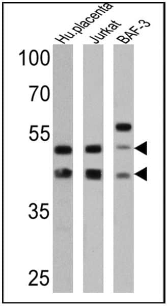
- Western blot analysis of CD47 was performed by loading 25 µg of human placenta (lane 1), Jurkat (lane 2) and BAF-3 (lane 3) cell lysates onto an SDS polyacrylamide gel. Proteins were transferred to a PVDF membrane and blocked at 4ºC overnight. The membrane was probed with a CD47 monoclonal antibody (Product # MA5-11895) at a dilution of 1:50 overnight at 4°C, washed in TBST, and probed with an HRP-conjugated secondary antibody for 1 hr at room temperature in the dark. Chemiluminescent detection was performed using Pierce ECL Plus Western Blotting Substrate (Product # 32132). Results show a band at ~42 and 52 kDa.
Supportive validation
- Submitted by
- Invitrogen Antibodies (provider)
- Main image
- Experimental details
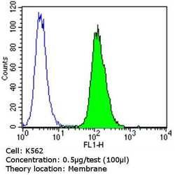
- Flow cytometry analysis of CD47 in K562 cells (green) compared to an isotype control (blue). Cells were harvested, adjusted to a concentration of 1-5x10^6 cells/mL, fixed with 2% paraformaldehyde and washed with PBS. Cells were blocked with a 2% solution of BSA-PBS for 30 min at room temperature and incubated with a CD47 monoclonal antibody (Product # MA5-11895) at a dilution of 0.5 µg/test for 60 min at room temperature. Cells were then incubated for 40 min at room temperature in the dark using a Dylight 488-conjugated goat anti-mouse IgG (H+L) secondary antibody and re-suspended in PBS for FACS analysis.

- Submitted by
- Invitrogen Antibodies (provider)
- Main image

- Experimental details
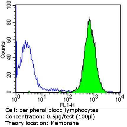
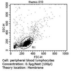
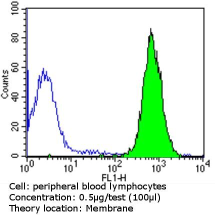
- Flow cytometry analysis of CD47 in PBMC cells (green) compared to an isotype control (blue). Human blood was collected, combined with a hydrophilic polysaccharide, centrifuged, transferred to a conical tube and washed with PBS. 50 µL of cell solution was added to each tube at a dilution of 2x10^7 cells/mL, followed by the addition of 50 µL of isotype control and primary antibody (Product # MA5-11895) at a dilution of 0.5 µg/test. Cells were incubated for 30 min at 4ºC and washed with a cell buffer, followed by incubation with a DyLight 488-conjugated goat anti-mouse IgG (H+L) secondary for 30 min at 4ºC in the dark. FACS analysis was performed using 400 µL of cell buffer.
- Submitted by
- Invitrogen Antibodies (provider)
- Main image
- Experimental details
- Flow cytometry analysis of CD47 in PBMC cells (green) compared to an isotype control (blue). Human blood was collected, combined with a hydrophilic polysaccharide, centrifuged, transferred to a conical tube and washed with PBS. 50 µL of cell solution was added to each tube at a dilution of 2x10^7 cells/mL, followed by the addition of 50 µL of isotype control and primary antibody (Product # MA5-11895) at a dilution of 0.5 µg/test. Cells were incubated for 30 min at 4ºC and washed with a cell buffer, followed by incubation with a DyLight 488-conjugated goat anti-mouse IgG (H+L) secondary for 30 min at 4ºC in the dark. FACS analysis was performed using 400 µL of cell buffer.

- Submitted by
- Invitrogen Antibodies (provider)
- Main image

- Experimental details
- Flow cytometry analysis of CD47 in PBMC cells (green) compared to an isotype control (blue). Human blood was collected, combined with a hydrophilic polysaccharide, centrifuged, transferred to a conical tube and washed with PBS. 50 µL of cell solution was added to each tube at a dilution of 2x10^7 cells/mL, followed by the addition of 50 µL of isotype control and primary antibody (Product # MA5-11895) at a dilution of 0.5 µg/test. Cells were incubated for 30 min at 4ºC and washed with a cell buffer, followed by incubation with a DyLight 488-conjugated goat anti-mouse IgG (H+L) secondary for 30 min at 4ºC in the dark. FACS analysis was performed using 400 µL of cell buffer.

Supportive validation
- Submitted by
- Invitrogen Antibodies (provider)
- Main image
- Experimental details
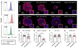
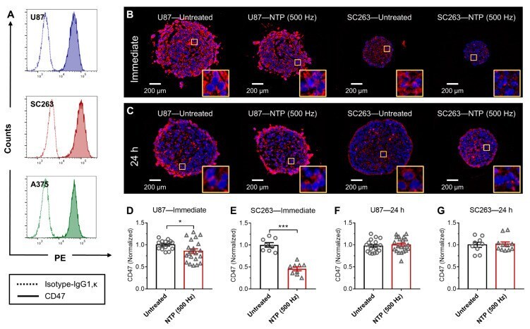
- Figure 1 NTP treatment modulated CD47 immediately in 3D tumor spheroids. ( A ) The baseline expression of three human cancer cells, glioblastoma (U87), head and neck squamous cell carcinoma (SC263), and melanoma (A375) was analyzed using immunohistochemistry and flow cytometry. U87 and SC263 cells were able to form compact spheroids and were exposed to NTP. Spheroids were collected ( B ) immediately or ( C ) 24 h after NTP treatment, paraffin-fixed, sectioned, stained for CD47 (red), and counter-stained with a nuclear dye, 4',6-diamidino-2-phenylindole (DAPI) (blue). Images were taken together at fixed microscope settings (10x) per experiment and all images were batch processed. Yellow inserts are a zoomed-in area (100 um x 100 um) to show CD47 staining surrounding the nucleus. CD47 expression was quantified and normalized to the untreated ( D - G ). Data here are represented as mean +- SEM and individual values are also shown (n = 8-21). Statistical significance of all treatment conditions was determined using the generalized linear mixed model with post hoc Dunnett's test comparison to untreated (* p
- Submitted by
- Invitrogen Antibodies (provider)
- Main image
- Experimental details
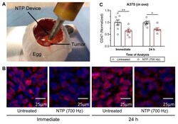
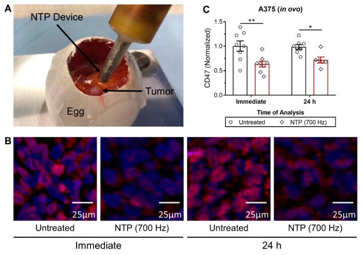
- Figure 2 NTP treatment decreased CD47 expression of A375 melanoma tumors in an in ovo model. ( A ) Tumors were grown on the chorioallantoic membrane (CAM) of fertilized eggs and treated directly with NTP. Following treatment, tumors were resected immediately or 24 h post treatment, sectioned, and ( B ) stained for CD47 (red) and counterstained with DAPI (blue). All images were taken on the same day at 10x and batch processed for quantification to limit variations. ( C ) Quantification of CD47 was normalized to the untreated. Data here are represented as mean +- SEM and individual values are also shown (n = 6-8). Statistical significance of all treatment conditions was determined using the generalized linear mixed model with post hoc Dunnett's test comparison to untreated (* p
- Submitted by
- Invitrogen Antibodies (provider)
- Main image
- Experimental details
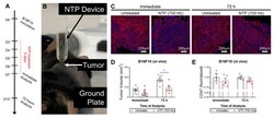
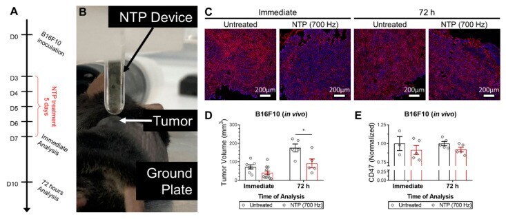
- Figure 3 NTP treatment reduced tumor volume and decreased CD47 expression, slightly, in vivo. ( A ) A schematic of the experimental design is provided. ( B ) B16F10 melanoma tumors were established in syngeneic B57BL/6 mice and treated directly with NTP for 5 consecutive days. Following treatment, tumors were resected immediately or 72 h post treatment and ( C ) stained for CD47 (red) and counterstained with DAPI (blue). All images were taken on the same day at 20x and batch processed for quantification to limit variations. ( D ) Tumor volumes were also reduced after treatment. ( E ) Quantification of CD47 was normalized to the untreated. Data here are represented as mean +- SEM and individual values are also shown (n = 3-11). Statistical significance of all treatment conditions was determined using the generalized linear mixed model with post hoc Dunnett's test comparison to untreated (* p
- Submitted by
- Invitrogen Antibodies (provider)
- Main image
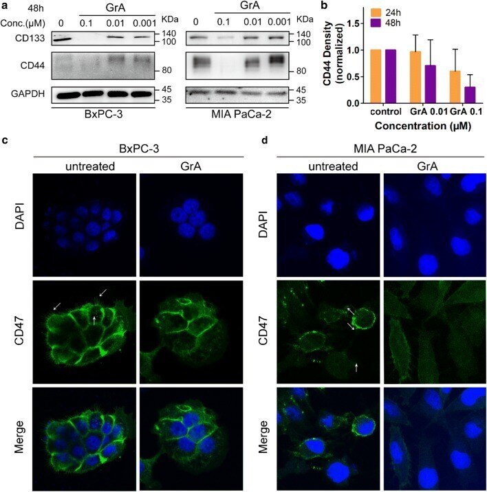
- Experimental details
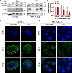
- Fig. 5 Effects of GrA on the expression and distribution of pancreatic CSC markers. a Protein expression profiles of CD133 and CD44 in BxPC-3 and MIA PaCa-2 cells treated with 0.1, 0.01 and 0.001 muM of GrA for 48 h represented graphically. b Densitometry quantitation of CD44 band intensities in western blot. Cells were treated with GrA for 24 h and 48 h, respectively. c Immunofluorescence staining images showed CD47 distribution in BxPC-3 cells treated with 0.05 muM GrA for 24 h. d Immunofluorescence staining images showed CD47 distribution in MIA PaCa-2 cells treated with 0.05 muM GrA for 24 h. Cellular nucleus was stained by DAPI exhibiting blue fluorescence while CD47 on cell surface was marked by antibodies with green fluorescence. The white arrow indicates the CD47 cluster