 Explore
 Validate
 Learn
Learn Western blot
Antibody data
- Antibody Data
- Antigen structure
- References [3]
- Comments [0]
- Validations
- Western blot [2]
- Immunohistochemistry [1]
- Flow cytometry [1]
- Blocking/Neutralizing [1]
Submit
Validation data
Reference
Comment
Report error
- Product number
- AF4670 - Provider product page

- Provider
- R&D Systems
- Product name
- Human CD47 Antibody
- Antibody type
- Polyclonal
- Description
- Antigen Affinity-purified. Detects human CD47 in direct ELISAs and Western blots. In direct ELISAs, less than 5% cross-reactivity with recombinant mouse CD47 is observed.
- Reactivity
- Human
- Host
- Sheep
- Conjugate
- Unconjugated
- Antigen sequence
Q08722- Isotype
- IgG
- Vial size
- 100 ug
- Concentration
- LYOPH
- Storage
- Use a manual defrost freezer and avoid repeated freeze-thaw cycles. 12 months from date of receipt, -20 to -70 °C as supplied. 1 month, 2 to 8 °C under sterile conditions after reconstitution. 6 months, -20 to -70 °C under sterile conditions after reconstitution.
Submitted references Combined prognostic value of the cancer stem cell markers CD47 and CD133 in esophageal squamous cell carcinoma.
Relationship between tumor-associated macrophage subsets and CD47 expression in squamous cell carcinoma of the head and neck in the tumor microenvironment.
Identification of a population of blood circulating tumor cells from breast cancer patients that initiates metastasis in a xenograft assay.
Wang JH, Huang ST, Zhang L, Liu ZG, Liang RX, Jiang SW, Jiang YN, Yu XJ, Jiang YC, Li XZ, Zhang PF, Wen ZS, Zheng M
Cancer medicine 2019 Mar;8(3):1315-1325
Cancer medicine 2019 Mar;8(3):1315-1325
Relationship between tumor-associated macrophage subsets and CD47 expression in squamous cell carcinoma of the head and neck in the tumor microenvironment.
Sakakura K, Takahashi H, Kaira K, Toyoda M, Murata T, Ohnishi H, Oyama T, Chikamatsu K
Laboratory investigation; a journal of technical methods and pathology 2016 Sep;96(9):994-1003
Laboratory investigation; a journal of technical methods and pathology 2016 Sep;96(9):994-1003
Identification of a population of blood circulating tumor cells from breast cancer patients that initiates metastasis in a xenograft assay.
Baccelli I, Schneeweiss A, Riethdorf S, Stenzinger A, Schillert A, Vogel V, Klein C, Saini M, Bäuerle T, Wallwiener M, Holland-Letz T, Höfner T, Sprick M, Scharpff M, Marmé F, Sinn HP, Pantel K, Weichert W, Trumpp A
Nature biotechnology 2013 Jun;31(6):539-44
Nature biotechnology 2013 Jun;31(6):539-44
No comments: Submit comment
Supportive validation
- Submitted by
- R&D Systems (provider)
- Main image
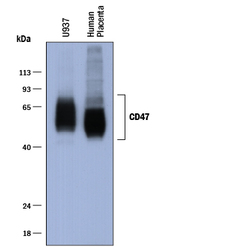
- Experimental details
- Detection of Human CD47 by Western Blot. fWestern blot shows lysates of U937 human histiocytic lymphoma cell line and human placenta tissue, not heated to minimize aggregation. PVDF membrane was probed with 1 µg/mL of Sheep Anti-Human CD47 Antigen Affinity-purified Polyclonal Antibody (Catalog # AF4670) followed by HRP-conjugated Anti-Sheep IgG Secondary Antibody (Catalog # HAF016). Specific bands were detected for CD47 at approximately 45-70 kDa (as indicated). This experiment was conducted under reducing conditions and using Immunoblot Buffer Group 1.
- Submitted by
- R&D Systems (provider)
- Main image
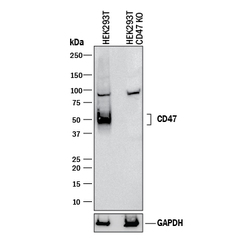
- Experimental details
- Western Blot Shows Human CD47 Specificity by Using Knockout Cell Line. Western blot shows lysates of HEK293T human embryonic kidney parental cell line and CD47 knockout HEK293T cell line (KO). PVDF membrane was probed with 1 µg/mL of Sheep Anti-Human CD47 Antigen Affinity-purified Polyclonal Antibody (Catalog # AF4670) followed by HRP-conjugated Anti-Sheep IgG Secondary Antibody (Catalog # HAF016). Specific bands were detected for CD47 at approximately 50 kDa (as indicated) in the parental HEK293T cell line, but is not detectable in knockout HEK293T cell line. GAPDH (Catalog # AF5718) is shown as a loading control. This experiment was conducted under reducing conditions and using Immunoblot Buffer Group 1.
Supportive validation
- Submitted by
- R&D Systems (provider)
- Main image
- Experimental details
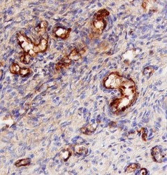
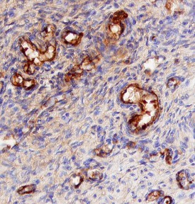
- CD47 in Human Placenta. CD47 was detected in immersion fixed paraffin-embedded sections of human placenta using 5 µg/mL Sheep Anti-Human CD47 Antigen Affinity-purified Polyclonal Antibody (Catalog # AF4670) overnight at 4 °C. Tissue was stained with the Anti-Sheep HRP-DAB Cell & Tissue Staining Kit (brown; Catalog # CTS019) and counterstained with hematoxylin (blue). View our protocol for Chromogenic IHC Staining of Paraffin-embedded Tissue Sections.
Supportive validation
- Submitted by
- R&D Systems (provider)
- Main image
- Experimental details
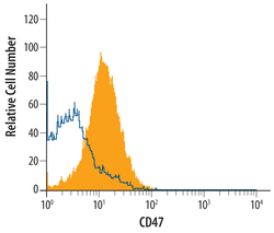
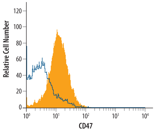
- Detection of CD47 in Human Lymphocytes by Flow Cytometry. Human whole blood lymphocytes were stained with Sheep Anti-Human CD47 Antigen Affinity-purified Polyclonal Antibody (Catalog # AF4670, filled histogram) or control antibody (Catalog # 5-001-A, open histogram), followed by Northern-Lights™ 557-conjugated Anti-Sheep IgG Secondary Antibody (Catalog # NL010).
Supportive validation
- Submitted by
- R&D Systems (provider)
- Main image
- Experimental details
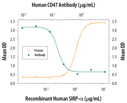
- Cell Adhesion Mediated by SIRP alpha/CD172a and Neutral-ization by Human CD47 Antibody. Recombinant Human SIRP alpha/CD172a (Catalog # 4546-SA), immobilized onto a microplate, supports the adhesion of the human erythrocytes in a dose-dependent manner (orange line). Adhesion elicited by Recombinant Human SIRP-alpha (2 µg/mL) is neutralized (green line) by increasing concentrations of Sheep Anti-Human CD47 Poly-clonal Antibody (Catalog # AF4670). The adhesion was maximally inhibited (70-100%) by 2 µg/mL of the antibody.
