Explore
 Validate
 Learn
Learn Western blot
 Immunocytochemistry
Antibody data
- Antibody Data
- Antigen structure
- References [2]
- Comments [0]
- Validations
- Immunocytochemistry [6]
- Chromatin Immunoprecipitation [3]
Submit
Validation data
Reference
Comment
Report error
- Product number
- PA5-17330 - Provider product page

- Provider
- Invitrogen Antibodies
- Product name
- AP2 gamma Polyclonal Antibody
- Antibody type
- Polyclonal
- Antigen
- Synthetic peptide
- Description
- It is not recommended to aliquot this antibody.
- Reactivity
- Human
- Host
- Rabbit
- Isotype
- IgG
- Vial size
- 100 μL
- Concentration
- 21 μg/mL
- Storage
- -20°C
Submitted references Fragile Gene WWOX Guides TFAP2A/TFAP2C-Dependent Actions Against Tumor Progression in Grade II Bladder Cancer.
WWOX Loses the Ability to Regulate Oncogenic AP-2γ and Synergizes with Tumor Suppressor AP-2α in High-Grade Bladder Cancer.
Kołat D, Kałuzińska Ż, Płuciennik E
Frontiers in oncology 2021;11:621060
Frontiers in oncology 2021;11:621060
WWOX Loses the Ability to Regulate Oncogenic AP-2γ and Synergizes with Tumor Suppressor AP-2α in High-Grade Bladder Cancer.
Kołat D, Kałuzińska Ż, Bednarek AK, Płuciennik E
Cancers 2021 Jun 12;13(12)
Cancers 2021 Jun 12;13(12)
No comments: Submit comment
Supportive validation
- Submitted by
- Invitrogen Antibodies (provider)
- Main image

- Experimental details
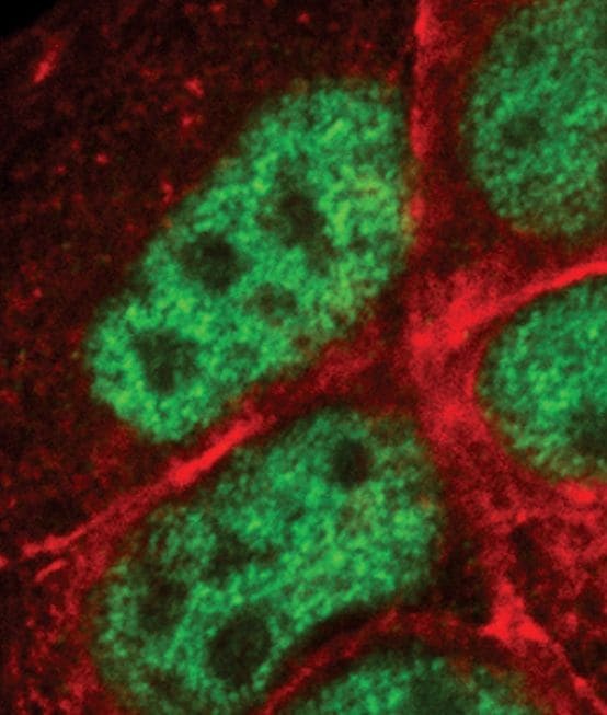
- Immunofluorescent analysis of AP-2-gamma in MCF-7 cells using an AP-2-gamma polyclonal antibody (Product # PA5-17330) (green). Actin filaments are labeled with a fluorescent red phalloidin.

- Submitted by
- Invitrogen Antibodies (provider)
- Main image

- Experimental details
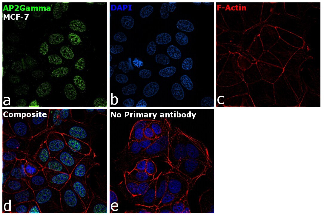
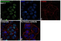
- Immunofluorescence analysis of AP2Gamma was performed using 70% confluent log phase MCF-7 cells. The cells were fixed with 4% paraformaldehyde for 10 minutes, permeabilized with 0.1% Triton™ X-100 for 15 minutes, and blocked with 2% BSA for 1 hour at room temperature. The cells were labeled with AP2 gamma Polyclonal Antibody (Product # PA5-17330) at 5 µg/mL in 0.1% BSA, incubated at 4 degree Celsius overnight and then labeled with Goat anti-Rabbit IgG (H+L) Superclonal™ Recombinant Secondary Antibody, Alexa Fluor® 488 conjugate (Product # A27034) at a dilution of 1:2000 for 45 minutes at room temperature (Panel a: green). Nuclei (Panel b: blue) were stained with SlowFade® Gold Antifade Mountant with DAPI (Product # S36938). F-actin (Panel c: red) was stained with Rhodamine Phalloidin (Product # R415, 1:300). Panel d represents the merged image showing localization to nucleus. Panel f represents control cells with no primary antibody to assess background. The images were captured at 60X magnification.
- Submitted by
- Invitrogen Antibodies (provider)
- Main image

- Experimental details
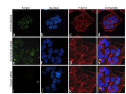
- Knockdown of AP2Gamma was achieved by transfecting MCF-7 cells with AP2Gamma specific siRNA (Silencer® select Product # s14010, s14009). Immunofluorescence analysis was performed on untransfected MCF-7 cells (panel a,d), transfected with non-specific scrambled siRNA (panels b,e) and transfected withAP2Gamma specific siRNA (panel c,f). Cells were fixed, permeabilized, and labelled with AP2 gamma Polyclonal Antibody (Product # PA5-17330), (1:100 dilution) followed by Goat anti-Rabbit IgG (H+L) Highly Cross-Adsorbed Secondary Antibody, Alexa Fluor Plus 488 (Product # A32723, 1:2000 dilution). Nuclei (blue) were stained with SlowFade® Gold Antifade Mountant with DAPI (Product # S36938), and Rhodamine Phalloidin (Product # R415, 1:300) was used for cytoskeletal F-actin (Red) staining. Partial reduction of specific signal was observed upon siRNA mediated knockdown (panel c,f) confirming specificity of the antibody to AP2Gamma (Green). The Images were captured at 60X magnification.

- Submitted by
- Invitrogen Antibodies (provider)
- Main image

- Experimental details
- Immunofluorescent analysis of AP-2-gamma in MCF-7 cells using an AP-2-gamma polyclonal antibody (Product # PA5-17330) (green). Actin filaments are labeled with a fluorescent red phalloidin.
- Submitted by
- Invitrogen Antibodies (provider)
- Main image
- Experimental details
- Immunofluorescence analysis of AP2Gamma was performed using 70% confluent log phase MCF-7 cells. The cells were fixed with 4% paraformaldehyde for 10 minutes, permeabilized with 0.1% Triton™ X-100 for 15 minutes, and blocked with 2% BSA for 1 hour at room temperature. The cells were labeled with AP2 gamma Polyclonal Antibody (Product # PA5-17330) at 5 µg/mL in 0.1% BSA, incubated at 4 degree Celsius overnight and then labeled with Goat anti-Rabbit IgG (Heavy Chain) Superclonal™ Recombinant Secondary Antibody, Alexa Fluor® 488 conjugate (Product # A27034) at a dilution of 1:2000 for 45 minutes at room temperature (Panel a: green). Nuclei (Panel b: blue) were stained with SlowFade® Gold Antifade Mountant with DAPI (Product # S36938). F-actin (Panel c: red) was stained with Rhodamine Phalloidin (Product # R415, 1:300). Panel d represents the merged image showing localization to nucleus. Panel f represents control cells with no primary antibody to assess background. The images were captured at 60X magnification.

- Submitted by
- Invitrogen Antibodies (provider)
- Main image
- Experimental details
- Knockdown of AP2Gamma was achieved by transfecting MCF-7 cells with AP2Gamma specific siRNA (Silencer® select Product # s14010, s14009). Immunofluorescence analysis was performed on untransfected MCF-7 cells (panel a,d), transfected with non-specific scrambled siRNA (panels b,e) and transfected withAP2Gamma specific siRNA (panel c,f). Cells were fixed, permeabilized, and labelled with AP2 gamma Polyclonal Antibody (Product # PA5-17330), (1:100 dilution) followed by Goat anti-Rabbit IgG (H+L) Highly Cross-Adsorbed Secondary Antibody, Alexa Fluor Plus 488 (Product # A32723, 1:2000 dilution). Nuclei (blue) were stained with SlowFade® Gold Antifade Mountant with DAPI (Product # S36938), and Rhodamine Phalloidin (Product # R415, 1:300) was used for cytoskeletal F-actin (Red) staining. Partial reduction of specific signal was observed upon siRNA mediated knockdown (panel c,f) confirming specificity of the antibody to AP2Gamma (Green). The Images were captured at 60X magnification.

Supportive validation
- Submitted by
- Invitrogen Antibodies (provider)
- Main image

- Experimental details
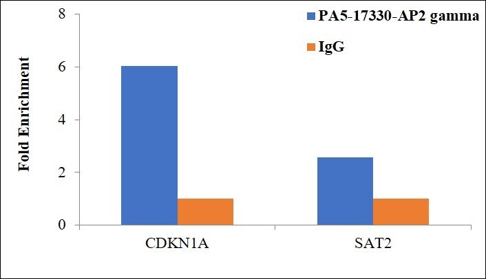
- Chromatin Immunoprecipitation (ChIP) assay of endogenous AP2 gamma protein using AP2 gamma Antibody: ChIP was performed using Anti-AP2 gamma Rabbit Polyclonal Antibody (Product # PA5-17330, 2.5 µg) on sheared chromatin from A431 cells using the MAGnify ChIP System kit (Product # 49-2024). Normal Rabbit IgG was used as a negative IP control. The purified DNA was analyzed by qPCR using primers binding to gene body of CDKN1A and SAT2 satellite repeats. Data is presented as fold enrichment of the antibody signal versus the negative control IgG using the comparative CT method.
- Submitted by
- Invitrogen Antibodies (provider)
- Main image
- Experimental details
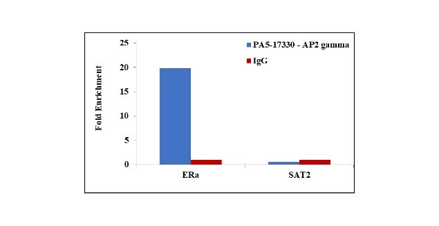
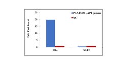
- Chromatin Immunoprecipitation (ChIP) assay of endogenous AP2 gamma protein using Anti-AP2 gamma Antibody: ChIP was performed using Anti-AP2 gamma Polyclonal Antibody (Product # PA5-17330, 2.5 µg) on sheared chromatin from MCF7 cells using the MAGnify ChIP System kit (Product # 49-2024). Normal Rabbit IgG was used as a negative IP control. The purified DNA was analyzed by qPCR using primers binding to ER-alpha promoter and SAT2 satellite repeats. Data is presented as fold enrichment of the antibody signal versus the negative control IgG using the comparative CT method.

- Submitted by
- Invitrogen Antibodies (provider)
- Main image

- Experimental details
- Chromatin Immunoprecipitation (ChIP) assay of endogenous AP2 gamma protein using Anti-AP2 gamma Antibody: ChIP was performed using Anti-AP2 gamma Polyclonal Antibody (Product # PA5-17330, 10 µl) on sheared chromatin from 2 million MCF7 cells using the MAGnify ChIP System kit (Product # 49-2024). Normal Rabbit IgG was used as a negative IP control. The purified DNA was analyzed by qPCR using primers binding to ER-alpha promoter and SAT2 satellite repeats. Data is presented as fold enrichment of the antibody signal versus the negative control IgG using the comparative CT method.