 Explore
 Validate
 Learn
Learn ELISA
ELISA Immunocytochemistry
Antibody data
- Antibody Data
- Antigen structure
- References [0]
- Comments [0]
- Validations
- Immunocytochemistry [1]
- Flow cytometry [1]
Submit
Validation data
Reference
Comment
Report error
- Product number
- MA5-14741 - Provider product page

- Provider
- Invitrogen Antibodies
- Product name
- Apolipoprotein B Monoclonal Antibody (F2C13)
- Antibody type
- Monoclonal
- Antigen
- Purifed from natural sources
- Description
- MA5-14741 targets Apolipoprotein B in ELISA, IP, and RIA applications and shows reactivity with Human samples. The MA5-14741 immunogen is purified human serum LDL. MA5-14741 detects Apolipoprotein B which has a predicted molecular weight of approximately 513 kDa. Product MA514741 is a smaller package size of MIA1610 (formerly sold as a Seradyn product).
- Reactivity
- Human
- Host
- Mouse
- Isotype
- IgG
- Antibody clone number
- F2C13
- Vial size
- 100 µg
- Concentration
- 1 mg/mL
- Storage
- Maintain refrigerated at 2-8°C for up to 6 months. For long term storage store at -20°C
No comments: Submit comment
Supportive validation
- Submitted by
- Invitrogen Antibodies (provider)
- Main image
- Experimental details
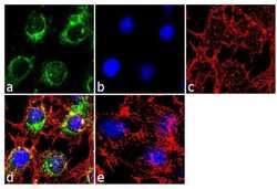
- Immunofluorescence analysis of Apolipoprotein B was performed using 70% confluent log phase Hep G2 cells. The cells were fixed with 4% paraformaldehyde for 10 minutes, permeabilized with 0.1% Triton™ X-100 for 10 minutes, and blocked with 2% BSA for 1 hour at room temperature. The cells were labeled with Apolipoprotein B (F2C13) Mouse Monoclonal Antibody (Product # MA5-14741) at 2 µg/mL in 0.1% BSA and incubated for 3 hours at room temperature and then labeled with Goat anti-Mouse IgG (H+L) Superclonal™ Secondary Antibody, Alexa Fluor® 488 conjugate (Product # A28175) a dilution of 1:2000 for 45 minutes at room temperature (Panel a: green). Nuclei (Panel b: blue) were stained with SlowFade® Gold Antifade Mountant with DAPI (Product # S36938). F-actin (Panel c: red) was stained with Alexa Fluor® 555 Rhodamine Phalloidin (Product # R415, 1:300). Panel d represents the merged image showing cytoplasmic localization. Panel e shows the no primary antibody control. The images were captured at 60X magnification.

Supportive validation
- Submitted by
- Invitrogen Antibodies (provider)
- Main image
- Experimental details
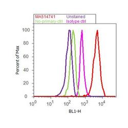
- Flow cytometry analysis of Apolipoprotein B was done on Hep G2 cells. Cells were fixed with 70% ethanol for 10 minutes, permeabilized with 0.25% Triton™ X-100 for 20 minutes, and blocked with 5% BSA for 30 minutes at room temperature. Cells were labeled with Apolipoprotein B Mouse Monoclonal Antibody (MA514741, red histogram) or with mouse isotype control (pink histogram) at 3-5 ug/million cells in 2.5% BSA. After incubation at room temperature for 2 hours, the cells were labeled with Alexa Fluor® 488 Rabbit Anti-Mouse Secondary Antibody (A11059) at a dilution of 1:400 for 30 minutes at room temperature. The representative 10,000 cells were acquired and analyzed for each sample using an Attune® Acoustic Focusing Cytometer. The purple histogram represents unstained control cells and the green histogram represents no-primary-antibody control..
