 Explore
 Validate
 Learn
Learn Western blot
 ELISA
ELISA Immunocytochemistry
Antibody data
- Antibody Data
- Antigen structure
- References [1]
- Comments [0]
- Validations
- Immunocytochemistry [3]
- Other assay [1]
Submit
Validation data
Reference
Comment
Report error
- Product number
- MIA1609 - Provider product page

- Provider
- Invitrogen Antibodies
- Product name
- Apolipoprotein B Monoclonal Antibody (F2C9)
- Antibody type
- Monoclonal
- Antigen
- Purifed from natural sources
- Description
- MIA1609 targets Apolipoprotein B in ELISA, IP and RIA applications and shows reactivity with Human samples. The MIA1609 immunogen is purified human serum LDL. MIA1609 detects Apolipoprotein B which has a predicted molecular weight of approximately 502 kDa. MIA1609 was formerly sold as a Seradyn product.
- Reactivity
- Human
- Host
- Mouse
- Isotype
- IgG
- Antibody clone number
- F2C9
- Vial size
- 1 mg
- Concentration
- 5 mg/mL
- Storage
- Maintain refrigerated at 2-8°C for up to 6 months. For long term storage store at -20°C
Submitted references Mass-Spectrometry Based Proteome Comparison of Extracellular Vesicle Isolation Methods: Comparison of ME-kit, Size-Exclusion Chromatography, and High-Speed Centrifugation.
Askeland A, Borup A, Østergaard O, Olsen JV, Lund SM, Christiansen G, Kristensen SR, Heegaard NHH, Pedersen S
Biomedicines 2020 Jul 25;8(8)
Biomedicines 2020 Jul 25;8(8)
No comments: Submit comment
Supportive validation
- Submitted by
- Invitrogen Antibodies (provider)
- Main image
- Experimental details
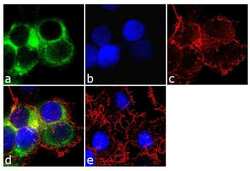
- Immunofluorescence analysis of Apolipoprotein B was performed using 70% confluent log phase Hep G2 cells. The cells were fixed with 4% paraformaldehyde for 10 minutes, permeabilized with 0.1% Triton™ X-100 for 10 minutes, and blocked with 2% BSA for 1 hour at room temperature. The cells were labeled with Apolipoprotein B (F2C9) Mouse Monoclonal Antibody (Product # MIA1609) at 2 µg/mL in 0.1% BSA and incubated for 3 hours at room temperature and then labeled with Goat anti-Mouse IgG (H+L) Superclonal™ Secondary Antibody, Alexa Fluor® 488 conjugate (Product # A28175) a dilution of 1:2000 for 45 minutes at room temperature (Panel a: green). Nuclei (Panel b: blue) were stained with SlowFade® Gold Antifade Mountant with DAPI (Product # S36938). F-actin (Panel c: red) was stained with Alexa Fluor® 555 Rhodamine Phalloidin (Product # R415, 1:300). Panel d represents the merged image showing cytoplasmic localization. Panel e shows the no primary antibody control. The images were captured at 60X magnification.

- Submitted by
- Invitrogen Antibodies (provider)
- Main image
- Experimental details
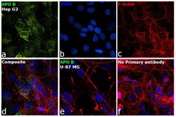
- Immunofluorescence analysis of Apolipoprotein B was performed using 70% confluent log phase Hep G2 cells. The cells were fixed with 4% paraformaldehyde for 5 minutes, permeabilized with 0.1% Triton™ X-100 for 10 minutes, and blocked with 2% BSA for 45 minutes at room temperature. The cells were labeled with Apolipoprotein B Monoclonal Antibody (F2C9) (Product # MIA1609) at 4 µg/mL concentration in 0.1% BSA, incubated at 4 degree celsius overnight and then labeled with Donkey anti-Mouse IgG (H+L) Highly Cross-Adsorbed Secondary Antibody, Alexa Fluor Plus 488 (Product # A32766), (1:2000 dilution), for 45 minutes at room temperature (Panel a: Green). Nuclei (Panel b: Blue) were stained with ProLong™ Diamond Antifade Mountant with DAPI (Product # P36962). F-actin (Panel c: Red) was stained with Rhodamine Phalloidin (Product # R415, 1:300). Panel d represents the merged image showing cytoplasm localization. Panel e represents negative cell line U-87 MG. Panel f represents control cells with no primary antibody to assess background. The images were captured at 60X magnification.

- Submitted by
- Invitrogen Antibodies (provider)
- Main image

- Experimental details
- Immunofluorescence analysis of Apolipoprotein B was performed using 70% confluent log phase Hep G2 cells. The cells were fixed with 4% paraformaldehyde for 5 minutes, permeabilized with 0.1% Triton™ X-100 for 10 minutes, and blocked with 2% BSA for 45 minutes at room temperature. The cells were labeled with Apolipoprotein B Monoclonal Antibody (F2C9) (Product # MIA1609) at 4 µg/mL concentration in 0.1% BSA, incubated at 4 degree celsius overnight and then labeled with Donkey anti-Mouse IgG (H+L) Highly Cross-Adsorbed Secondary Antibody, Alexa Fluor Plus 488 (Product # A32766), (1:2000 dilution), for 45 minutes at room temperature (Panel a: Green). Nuclei (Panel b: Blue) were stained with ProLong™ Diamond Antifade Mountant with DAPI (Product # P36962). F-actin (Panel c: Red) was stained with Rhodamine Phalloidin (Product # R415, 1:300). Panel d represents the merged image showing cytoplasm localization. Panel e represents negative cell line U-87 MG. Panel f represents control cells with no primary antibody to assess background. The images were captured at 60X magnification.

Supportive validation
- Submitted by
- Invitrogen Antibodies (provider)
- Main image
- Experimental details
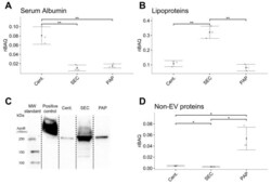
- Figure 5 Difference in the co-isolation of contaminants such as serum albumin, lipoproteins, and other non-EV proteins between isolates produced by high-speed centrifugation (Cent. ), size-exclusion chromatography (SEC), and peptide affinity precipitation (PAP). ( A ) High-speed centrifugation produced EV isolates with the highest abundance of serum albumin. ( B ) SEC isolates contained the highest amount of lipoproteins. ( C ) Immunoblotting confirms elevated levels of apolipoprotein B in isolates produced by SEC. ( D ) PAP isolates contained the highest amount of non-EV proteins. Error bars: Mean +- SD. Significance levels: * ( p -value < 0.05) and ** ( p -value < 0.01).
