 Explore
 Validate
 Learn
LearnMA1-16675
antibody from Invitrogen Antibodies
Targeting: CELF1
BRUNOL2, CUG-BP, CUGBP, CUGBP1, EDEN-BP, hNab50, NAB50, NAPOR
 Western blot
Antibody data
- Antibody Data
- Antigen structure
- References [1]
- Comments [0]
- Validations
- Western blot [2]
- Immunocytochemistry [2]
- Immunohistochemistry [1]
- Flow cytometry [2]
Submit
Validation data
Reference
Comment
Report error
- Product number
- MA1-16675 - Provider product page

- Provider
- Invitrogen Antibodies
- Product name
- CUGBP1 Monoclonal Antibody (3B1)
- Antibody type
- Monoclonal
- Antigen
- Other
- Description
- Suggested positive control: antigen standard for CUGBP1 (transient overexpression lysate).
- Reactivity
- Human, Mouse, Rat, Bovine, Porcine, Rabbit
- Host
- Mouse
- Isotype
- IgG
- Antibody clone number
- 3B1
- Vial size
- 100 μL
- Concentration
- 1 mg/mL
- Storage
- -20°C, Avoid Freeze/Thaw Cycles
Submitted references Analysis of MTMR1 expression and correlation with muscle pathological features in juvenile/adult onset myotonic dystrophy type 1 (DM1) and in myotonic dystrophy type 2 (DM2).
Santoro M, Modoni A, Masciullo M, Gidaro T, Broccolini A, Ricci E, Tonali PA, Silvestri G
Experimental and molecular pathology 2010 Oct;89(2):158-68
Experimental and molecular pathology 2010 Oct;89(2):158-68
No comments: Submit comment
Supportive validation
- Submitted by
- Invitrogen Antibodies (provider)
- Main image

- Experimental details
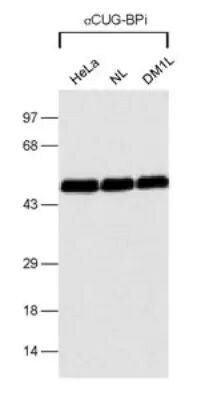
- Western blot analysis of CUGBP1 in several cell lysates. Sample was incubated in CUGBP1 monoclonal antibody (Product # MA1-16675).
- Submitted by
- Invitrogen Antibodies (provider)
- Main image

- Experimental details
- Western blot analysis of CUGBP1 in 0.5 mg/mL HeLa lysate. Samples were incubated in CUGBP1 monoclonal antibody (Product # MA1-16675). This experiment was performed under reducing conditions using the 12-230 kDa separation system..

Supportive validation
- Submitted by
- Invitrogen Antibodies (provider)
- Main image

- Experimental details
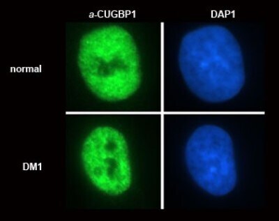
- Immunocytochemistry analysis of CUGBP1 in normal and DM1 (dystrophia myotonica) myoblasts. Samples were incubated in CUGBP1 monoclonal antibody (Product # MA1-16675). Subcellular distribution of CUGBP1 (nuclear, non-nucleolar).
- Submitted by
- Invitrogen Antibodies (provider)
- Main image

- Experimental details
- Immunocytochemistry analysis of CUGBP1 in normal and DM1 (dystrophia myotonica) myoblasts. Samples were incubated in CUGBP1 monoclonal antibody (Product # MA1-16675). Subcellular distribution of CUGBP1 (nuclear, non-nucleolar).

Supportive validation
- Submitted by
- Invitrogen Antibodies (provider)
- Main image
- Experimental details
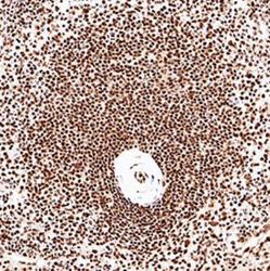
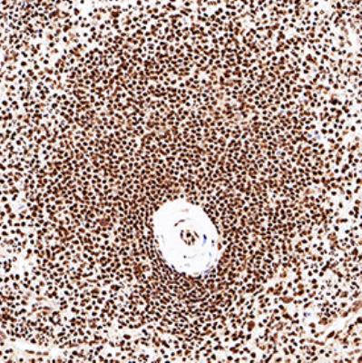
- Immunohistochemical analysis of CUGBP1 in formalin fixed paraffin-embedded (FFPE) human spleen. Samples were incubated in CUGBP1 monoclonal antibody (Product # MA1-16675) using a dilution of 1:100. Bond Rx autostainer (Leica Biosystems). The assay involved 30 minutes of heat induced antigen retrieval (HIER) using 10mM sodium citrate buffer (pH 9.0) and endogenous peroxidase quenching with peroxide block. The sections were incubated with primary antibody for 15 minutes and Bond Polymer Refine Detection (Leica Biosystems) with DAB was used for signal development followed by counterstaining with hematoxylin. Nuclear staining was observed in lymphocytes.
Supportive validation
- Submitted by
- Invitrogen Antibodies (provider)
- Main image

- Experimental details
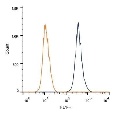
- Flow cytometry of CUGBP1 in 1 x 10^6 MCF-7 cells. Samples were incubated in CUGBP1 monoclonal antibody (Product # MA1-16675) using a dilution of 1 µg/1x10^6 cells. Antibody (dark blue). Isotype control shown in orange.
- Submitted by
- Invitrogen Antibodies (provider)
- Main image
- Experimental details
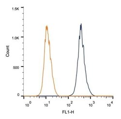
- Flow cytometry of CUGBP1 in 1 x 10^6 MCF-7 cells. Samples were incubated in CUGBP1 monoclonal antibody (Product # MA1-16675) using a dilution of 1 µg/1x10^6 cells. Antibody (dark blue). Isotype control shown in orange.
