Explore
 Validate
 Learn
Learn Western blot
 ELISA
ELISA Immunocytochemistry
Antibody data
- Antibody Data
- Antigen structure
- References [2]
- Comments [0]
- Validations
- Immunocytochemistry [2]
- Immunohistochemistry [2]
- Flow cytometry [2]
- Other assay [2]
Submit
Validation data
Reference
Comment
Report error
- Product number
- MA5-17083 - Provider product page

- Provider
- Invitrogen Antibodies
- Product name
- Glypican 3 Monoclonal Antibody (9C2)
- Antibody type
- Monoclonal
- Antigen
- Purifed from natural sources
- Description
- MA5-17083 targets GPC3 in indirect ELISA, FACS, ICC, IHC, IF and WB applications and shows reactivity with Human and Mouse samples. The MA5-17083 immunogen is purified recombinant fragment of human GPC3 expressed in E. Coli. MA5-17083 detects GPC3 which has a predicted molecular weight of approximately 65.5kDa.
- Reactivity
- Human, Mouse
- Host
- Mouse
- Isotype
- IgG
- Antibody clone number
- 9C2
- Vial size
- 100 μg
- Concentration
- 1 mg/mL
- Storage
- Store at 4°C short term. For long term storage, store at -20°C, avoiding freeze/thaw cycles.
Submitted references Identification of miR-4510 as a metastasis suppressor of gastric cancer through regulation of tumor microenvironment via targeting GPC3.
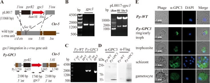
Plasmodium parasite as an effective hepatocellular carcinoma antigen glypican-3 delivery vector.
Ma HF, Shu P, Shi XH, Wang M, Jiang MF
Clinical & experimental metastasis 2022 Apr;39(2):363-374
Clinical & experimental metastasis 2022 Apr;39(2):363-374
Plasmodium parasite as an effective hepatocellular carcinoma antigen glypican-3 delivery vector.
Liu Q, Yang Y, Tan X, Tao Z, Adah D, Yu S, Lu J, Zhao S, Qin L, Qin L, Chen X
Oncotarget 2017 Apr 11;8(15):24785-24796
Oncotarget 2017 Apr 11;8(15):24785-24796
No comments: Submit comment
Supportive validation
- Submitted by
- Invitrogen Antibodies (provider)
- Main image

- Experimental details
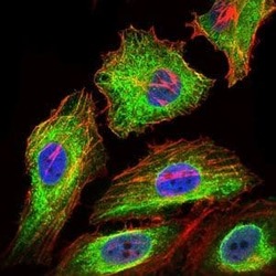
- Immunofluorescence analysis of HeLa cells using GPC3 monoclonal antibody (Product # MA5-17083) (Green). Blue: DRAQ5 fluorescent DNA dye. Red: actin filaments have been labeled with phalloidin.

- Submitted by
- Invitrogen Antibodies (provider)
- Main image
- Experimental details
- Immunofluorescence analysis of HeLa cells using GPC3 monoclonal antibody (Product # MA5-17083) (Green). Blue: DRAQ5 fluorescent DNA dye. Red: actin filaments have been labeled with phalloidin.

Supportive validation
- Submitted by
- Invitrogen Antibodies (provider)
- Main image
- Experimental details
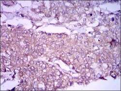
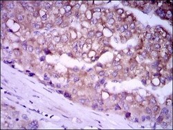
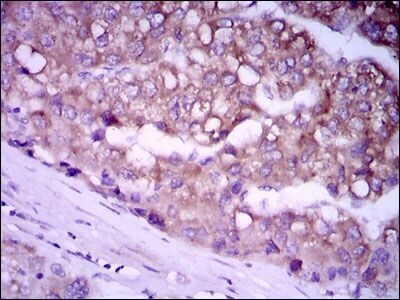
- Immunohistochemical analysis of paraffin-embedded breast cancer tissues using GPC3 monoclonal antibody (Product # MA5-17083) followed with DAB staining.

- Submitted by
- Invitrogen Antibodies (provider)
- Main image
- Experimental details
- Immunohistochemical analysis of paraffin-embedded liver cancer tissues using GPC3 monoclonal antibody (Product # MA5-17083) followed with DAB staining.
Supportive validation
- Submitted by
- Invitrogen Antibodies (provider)
- Main image

- Experimental details
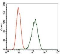
- Flow cytometric analysis of Jurkat cells using GPC3 monoclonal antibody (Product # MA5-17083) (green) and negative control (red).
- Submitted by
- Invitrogen Antibodies (provider)
- Main image

- Experimental details
- Flow cytometric analysis of Jurkat cells using GPC3 monoclonal antibody (Product # MA5-17083) (green) and negative control (red).

Supportive validation
- Submitted by
- Invitrogen Antibodies (provider)
- Main image
- Experimental details
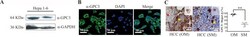
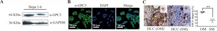
- Figure 1 GPC3 protein is highly expressed in Hepa1-6 cells and Hepa1-6 cell-induced HCC tissues in mice ( A ) Western blot analysis of GPC3 protein in Hepa1-6 cells. ( B ) Localization of GPC3 in Hepa1-6 cells using confocal microscopy. ( C ) Immunohistochemical analysis of GPC3 in the HCC tissues. The above data is a representation of four repeated experiments. Scatter plot show the mean percentage +- SD of GPC3 positive Hepa1-6 cells. Statistical differences between groups are indicated by the P values (* P
- Submitted by
- Invitrogen Antibodies (provider)
- Main image
- Experimental details
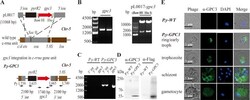
- Figure 2 Expression of ectopic GPC3 by stably transfected P. yoelii 17XNL ( A ) Schematic representation of the pL0017- gpc3 vector integrated into the c-rrna unit. ( B ) Gel electrophoresis and DNA analysis of the gpc3 cloned from cDNAs of Hepa1-6 cells (left) by PCR, and identification of the pL0017- gpc3 vector (right) by restriction enzyme analysis ( Bam HI/ Xba I). ( C ) Correct integration of the vector in P.y -GPC3. PCR was performed with genomic DNA from P.y-GPC3 and P.y-WT (negative control). The 5' integration site ( 5' int , primers PyL739/L635) and 3' integration site ( 3' int , primers L665/PyL740) were verified, as well as the gpc3 gene. ( D ) Detection of GPC3 expression in P.y-GPC3 by western blot using anti-GPC3 (rabbit) or anti-Flag tag antibodies. ( E ) GPC3 expression by the four different stages of the P.y-GPC3 . Parasitized erythrocytes were analyzed by immunofluorescence and P.y-WT infected erythrocytes were used for the negative control. Parasites stained with anti-GPC3 antibody (green), or DAPI for DNA, were visualized by confocal microscopy. Bars: 5 mum.