Explore
 Validate
 Learn
Learn Western blot
 Immunohistochemistry
 Flow cytometry
Antibody data
- Antibody Data
- Antigen structure
- References [1]
- Comments [0]
- Validations
- Western blot [3]
- ELISA [1]
- Immunocytochemistry [1]
Submit
Validation data
Reference
Comment
Report error
- Product number
- PA5-47256 - Provider product page

- Provider
- Invitrogen Antibodies
- Product name
- Glypican 3 Polyclonal Antibody
- Antibody type
- Polyclonal
- Antigen
- Recombinant full-length protein
- Description
- In direct ELISAs, less than 10% cross-reactivity with recombinant mouse Glypican 3 is observed and less than 5% cross-reactivity with recombinant human (rh) Glypican 2, rhGlypican 5, and rhGlypican 6 is observed. Reconstitute at 0.2 mg/mL in sterile PBS.
- Reactivity
- Human
- Host
- Sheep
- Isotype
- IgG
- Vial size
- 100 µg
- Concentration
- 0.2 mg/mL
- Storage
- -20° C, Avoid Freeze/Thaw Cycles
Submitted references Transferrin Receptor Is a Specific Ferroptosis Marker.
Feng H, Schorpp K, Jin J, Yozwiak CE, Hoffstrom BG, Decker AM, Rajbhandari P, Stokes ME, Bender HG, Csuka JM, Upadhyayula PS, Canoll P, Uchida K, Soni RK, Hadian K, Stockwell BR
Cell reports 2020 Mar 10;30(10):3411-3423.e7
Cell reports 2020 Mar 10;30(10):3411-3423.e7
No comments: Submit comment
Supportive validation
- Submitted by
- Invitrogen Antibodies (provider)
- Main image

- Experimental details
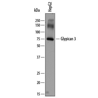
- Western blot analysis from lysates of HepG2 human hepatocellular carcinoma cell line. PVDF membrane was probed with 2 µg/mL of Sheep Anti-human Glypican 3 Antigen Affinity-purified Polyclonal Antibody (Product # PA5-47256) followed by HRP-conjugated Anti-Sheep IgG Secondary Antibody. A specific band was detected for Glypican 3 at approximately 75 kDa (as indicated). This experiment was conducted under reducing conditions.

- Submitted by
- Invitrogen Antibodies (provider)
- Main image

- Experimental details
- Western blot analysis of Glypican 3 in HepG2 human hepatocellular carcinoma cell line. Samples were incubated in Glypican 3 polyclonal antibody (Product # PA5-47256) using a dilution of 2 µg/mL followed by a HRP-conjugated Anti-Sheep IgG secondary antibody. A specific band was detected for Glypican 3 at approximately 75 kDa (as indicated). This experiment was conducted under reducing conditions.
- Submitted by
- Invitrogen Antibodies (provider)
- Main image
- Experimental details
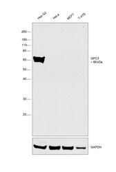
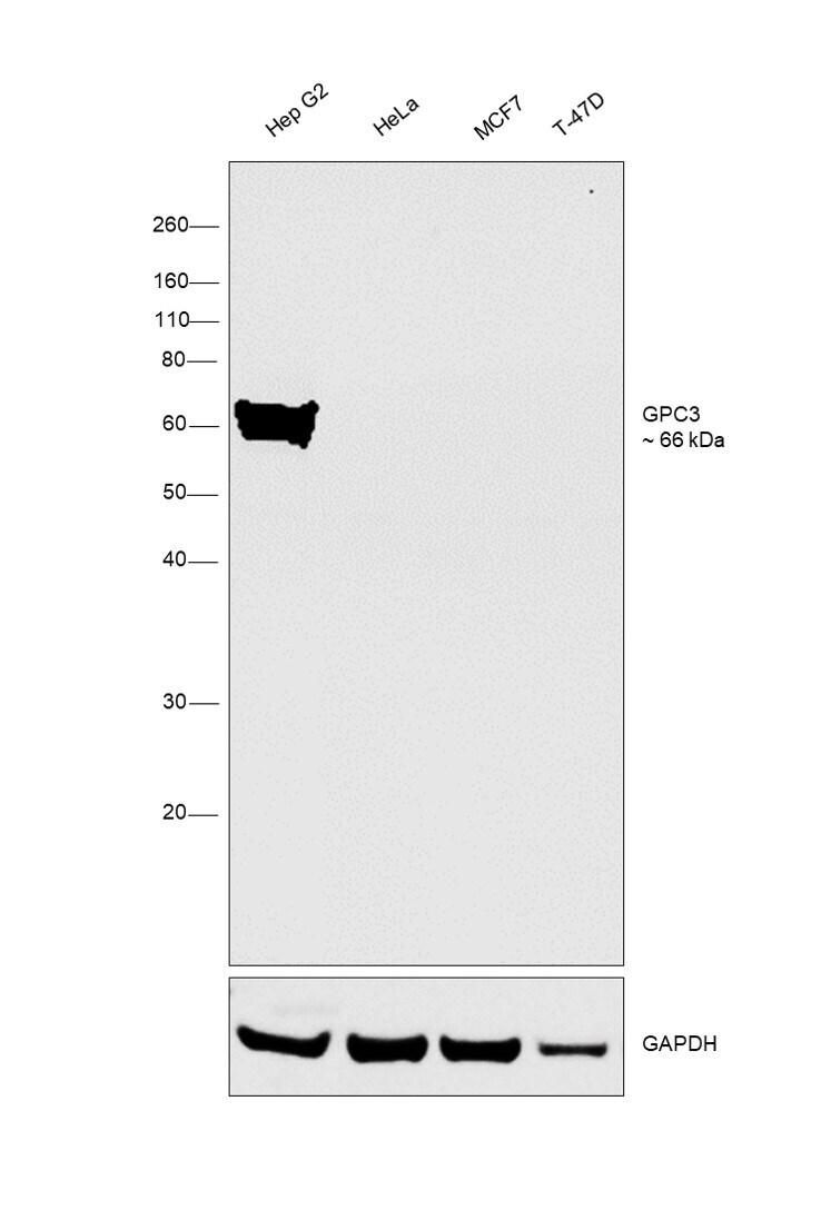
- Western blot was performed using Glypican 3 Polyclonal Antibody (Product # PA5-47256) and a 66 kDa band corresponding to GPC3 was observed only in Hep G2. Membrane enriched extracts (30 µg lysate) of Hep G2 (Lane 1), HeLa (Lane 2), MCF7 (Lane 3) and T-47D (Lane 4) were electrophoresed using NuPAGE™ 10% Bis-Tris Protein Gel (Product # NP0302BOX). Resolved proteins were then transferred onto a nitrocellulose membrane (Product # IB23001) by iBlot® 2 Dry Blotting System (Product # IB21001). The blot was probed with the primary antibody (1:500 dilution) and detected by chemiluminescence with Rabbit anti-Sheep IgG (H+L), Secondary Antibody, HRP (Product # 61-8620, 1:4000 dilution) using the iBright FL 1000 (Product # A32752). Chemiluminescent detection was performed using Novex® ECL Chemiluminescent Substrate Reagent Kit (Product # WP20005).
Supportive validation
- Submitted by
- Invitrogen Antibodies (provider)
- Main image

- Experimental details
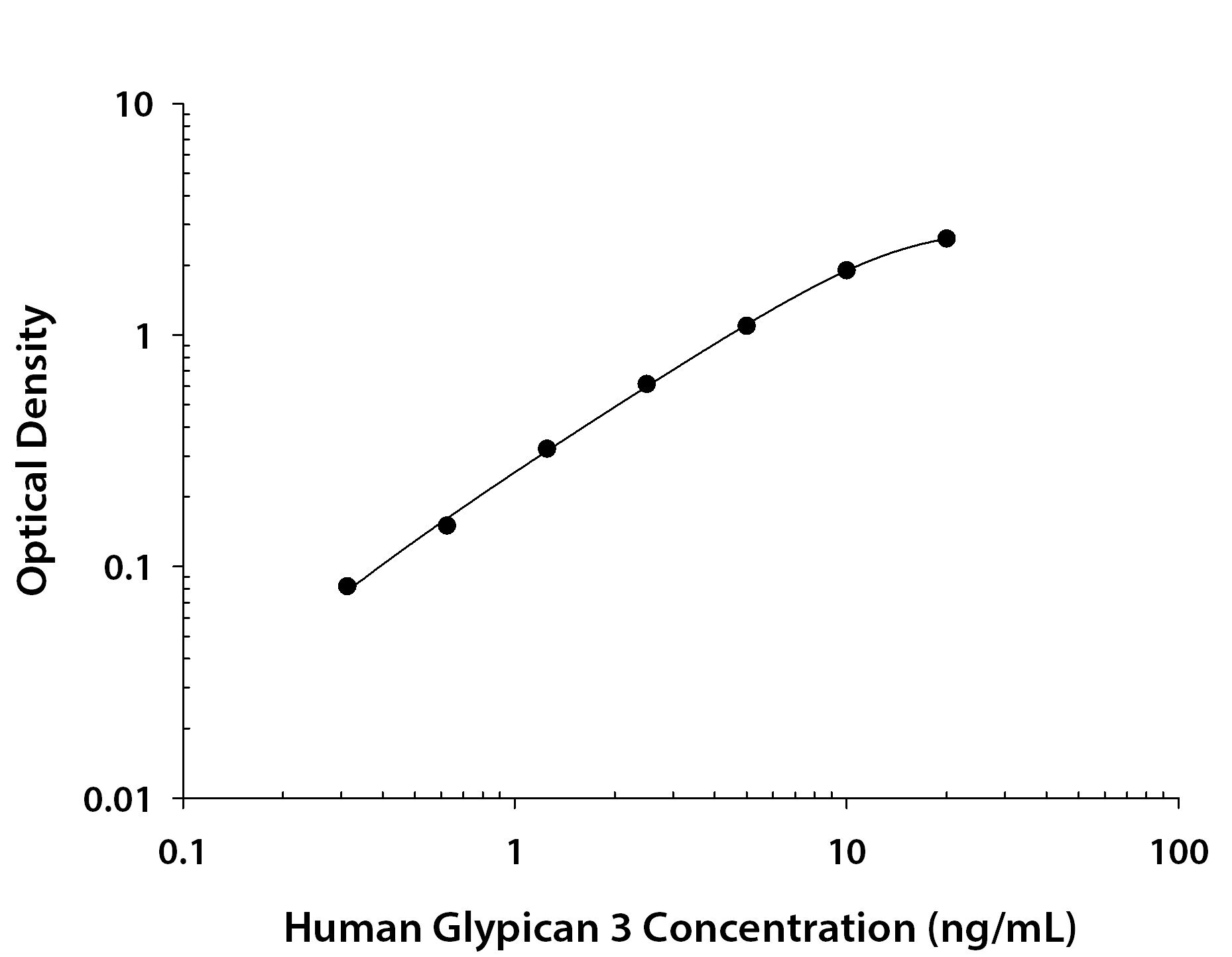
- ELISA of Glypican 3 in recombinant Glypican 3 protein. Samples were incubated in Glypican 3 polyclonal antibody (Product # PA5-47256). Protein was serially diluted 2-fold. Antibody was biotinylated and incubated with the protein captured on the plate. Detection of the standard curve was achieved by incubating Streptavidin-HRP followed by Substrate Solution and stopping the enzymatic reaction with Stop Solution.
Supportive validation
- Submitted by
- Invitrogen Antibodies (provider)
- Main image

- Experimental details
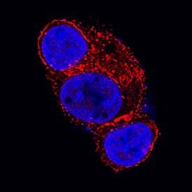
- Immunocytochemistry analysis of Glypican 3 in immersion fixed HepG2 human hepatocellular carcinoma cell line. Samples were incubated in Glypican 3 polyclonal antibody (Product # PA5-47256) using a dilution of 1.7 µg/mL for 3 hours at room temperature followed by NorthernLights™ 557-conjugated Anti-Sheep IgG Secondary Antibody (red) and counterstained with DAPI (blue). Specific staining was localized to cytoplasm.