Explore
 Validate
 Learn
Learn Western blot
 Flow cytometry
Antibody data
- Antibody Data
- Antigen structure
- References [3]
- Comments [0]
- Validations
- Western blot [1]
- Immunocytochemistry [1]
- Immunohistochemistry [2]
Submit
Validation data
Reference
Comment
Report error
- Product number
- AF2119 - Provider product page

- Provider
- R&D Systems
- Product name
- Human Glypican 3 Antibody
- Antibody type
- Polyclonal
- Description
- Antigen Affinity-purified. Detects human Glypican 3 in direct ELISAs and Western blots. In direct ELISAs, less than 10% cross-reactivity with recombinant mouse Glypican 3 is observed and less than 5% cross-reactivity with recombinant human (rh) Glypican 2, rhGlypican 5, and rhGlypican 6 is observed.
- Reactivity
- Human
- Host
- Sheep
- Conjugate
- Unconjugated
- Antigen sequence
P51654- Isotype
- IgG
- Vial size
- 100 ug
- Concentration
- LYOPH
- Storage
- Use a manual defrost freezer and avoid repeated freeze-thaw cycles. 12 months from date of receipt, -20 to -70 °C as supplied. 1 month, 2 to 8 °C under sterile conditions after reconstitution. 6 months, -20 to -70 °C under sterile conditions after reconstitution.
Submitted references Phase I trial of a glypican-3-derived peptide vaccine for advanced hepatocellular carcinoma: immunologic evidence and potential for improving overall survival.
Bone-specific heparan sulfates induce osteoblast growth arrest and downregulation of retinoblastoma protein.
The oncofetal protein glypican-3 is a novel marker of hepatic progenitor/oval cells.
Sawada Y, Yoshikawa T, Nobuoka D, Shirakawa H, Kuronuma T, Motomura Y, Mizuno S, Ishii H, Nakachi K, Konishi M, Nakagohri T, Takahashi S, Gotohda N, Takayama T, Yamao K, Uesaka K, Furuse J, Kinoshita T, Nakatsura T
Clinical cancer research : an official journal of the American Association for Cancer Research 2012 Jul 1;18(13):3686-96
Clinical cancer research : an official journal of the American Association for Cancer Research 2012 Jul 1;18(13):3686-96
Bone-specific heparan sulfates induce osteoblast growth arrest and downregulation of retinoblastoma protein.
Manton KJ, Sadasivam M, Cool SM, Nurcombe V
Journal of cellular physiology 2006 Oct;209(1):219-29
Journal of cellular physiology 2006 Oct;209(1):219-29
The oncofetal protein glypican-3 is a novel marker of hepatic progenitor/oval cells.
Grozdanov PN, Yovchev MI, Dabeva MD
Laboratory investigation; a journal of technical methods and pathology 2006 Dec;86(12):1272-84
Laboratory investigation; a journal of technical methods and pathology 2006 Dec;86(12):1272-84
No comments: Submit comment
Supportive validation
- Submitted by
- R&D Systems (provider)
- Main image
- Experimental details
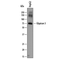
- Detection of Human Glypican 3 by Western Blot. Western blot shows lysates of HepG2 human hepatocellular carcinoma cell line. PVDF membrane was probed with 2 µg/mL of Sheep Anti-Human Glypican 3 Antigen Affinity-purified Polyclonal Antibody (Catalog # AF2119) followed by HRP-conjugated Anti-Sheep IgG Secondary Antibody (Catalog # HAF016). A specific band was detected for Glypican 3 at approximately 75 kDa (as indicated). This experiment was conducted under reducing conditions and using Immunoblot Buffer Group 1.

Supportive validation
- Submitted by
- R&D Systems (provider)
- Main image

- Experimental details
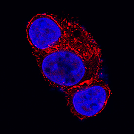
- Glypican 3 in HepG2 Human Cell Line. Glypican 3 was detected in immersion fixed HepG2 human hepatocellular carcinoma cell line using Sheep Anti-Human Glypican 3 Antigen Affinity-purified Polyclonal Antibody (Catalog # AF2119) at 1.7 µg/mL for 3 hours at room temperature. Cells were stained using the NorthernLights™ 557-conjugated Anti-Sheep IgG Secondary Antibody (red; Catalog # NL010) and counterstained with DAPI (blue). Specific staining was localized to cytoplasm and cell membranes. View our protocol for Fluorescent ICC Staining of Cells on Coverslips.
Supportive validation
- Submitted by
- R&D Systems (provider)
- Main image

- Experimental details
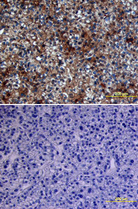
- Glypican 3 in Human Breast. Glypican 3 was detected in immersion fixed paraffin-embedded sections of human breast using 5 µg/mL Sheep Anti-Human Glypican 3 Antigen Affinity-purified Polyclonal Antibody (Catalog # AF2119) overnight at 4 °C. Tissue was stained with the Anti-Sheep HRP-DAB Cell & Tissue Staining Kit (brown; Catalog # CTS019) and counterstained with hematoxylin (blue). View our protocol for Chromogenic IHC Staining of Paraffin-embedded Tissue Sections.
- Submitted by
- R&D Systems (provider)
- Main image
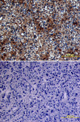
- Experimental details
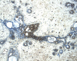
- Glypican 3 in Human Liver Cancer Tissue. Glypican 3 was detected in immersion fixed paraffin-embedded sections of human liver cancer tissue using Sheep Anti-Human Glypican 3 Antigen Affinity-purified Polyclonal Antibody (Catalog # AF2119) at 10 µg/mL overnight at 4 °C. Tissue was stained using the Anti-Sheep HRP-DAB Cell & Tissue Staining Kit (brown; Catalog # CTS019) and counterstained with hematoxylin (blue). Lower panel shows a lack of labeling if primary antibodies are omitted and tissue is stained only with secondary antibody followed by incubation with detection reagents. View our protocol for Chromogenic IHC Staining of Paraffin-embedded Tissue Sections.