Explore
 Validate
 Learn
Learn Western blot
 Flow cytometry
Antibody data
- Antibody Data
- Antigen structure
- References [2]
- Comments [0]
- Validations
- Western blot [2]
- Immunocytochemistry [1]
Submit
Validation data
Reference
Comment
Report error
- Product number
- MAB2119 - Provider product page

- Provider
- R&D Systems
- Product name
- Human/Mouse/Rat Glypican 3 Antibody
- Antibody type
- Monoclonal
- Description
- Protein A or G purified from hybridoma culture supernatant. Detects human Glypican 3 in ELISAs. Deteacts human, mouse, and rat Glypican 3 in Western blots. Does not cross-react with recombinant human (rh) Glypican-2, rhGlypican-5, or rhGlypican-6.
- Reactivity
- Human, Mouse, Rat
- Host
- Mouse
- Conjugate
- Unconjugated
- Antigen sequence
P51654.1- Isotype
- IgG
- Antibody clone number
- 307801
- Vial size
- 100 ug
- Concentration
- LYOPH
- Storage
- Use a manual defrost freezer and avoid repeated freeze-thaw cycles. 12 months from date of receipt, -20 to -70 °C as supplied. 1 month, 2 to 8 °C under sterile conditions after reconstitution. 6 months, -20 to -70 °C under sterile conditions after reconstitution.
Submitted references Autophagy suppresses proliferation of HepG2 cells via inhibiting glypican-3/wnt/β-catenin signaling.
The heparan sulfate proteoglycan (HSPG) glypican-3 mediates commitment of MC3T3-E1 cells toward osteogenesis.
Hu P, Cheng B, He Y, Wei Z, Wu D, Meng Z
OncoTargets and therapy 2018;11:193-200
OncoTargets and therapy 2018;11:193-200
The heparan sulfate proteoglycan (HSPG) glypican-3 mediates commitment of MC3T3-E1 cells toward osteogenesis.
Haupt LM, Murali S, Mun FK, Teplyuk N, Mei LF, Stein GS, van Wijnen AJ, Nurcombe V, Cool SM
Journal of cellular physiology 2009 Sep;220(3):780-91
Journal of cellular physiology 2009 Sep;220(3):780-91
No comments: Submit comment
Supportive validation
- Submitted by
- R&D Systems (provider)
- Main image
- Experimental details
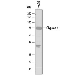
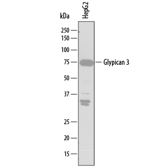
- Detection of Human Glypican 3 by Western Blot. Western blot shows lysates of HepG2 human hepatocellular carcinoma cell line. PVDF membrane was probed with 2 µg/mL of Mouse Anti-Human Glypican 3 Monoclonal Antibody (Catalog # MAB2119) followed by HRP-conjugated Anti-Mouse IgG Secondary Antibody (Catalog # HAF007). A specific band was detected for Glypican 3 at approximately 75 kDa (as indicated). This experiment was conducted under reducing conditions and using Immunoblot Buffer Group 1.
- Submitted by
- R&D Systems (provider)
- Main image
- Experimental details
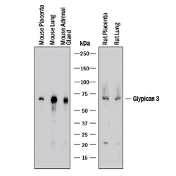
- Detection of Mouse and Rat Glypican 3 by Western Blot. Western blot shows lysates of mouse placenta tissue, mouse lung tissue, mouse adrenal gland tissue, rat placenta tissue, and rat lung tissue. PVDF membrane was probed with 1 µg/mL of Mouse Anti-Human Glypican 3 Monoclonal Antibody (Catalog # MAB2119) followed by HRP-conjugated Anti-Mouse IgG Secondary Antibody (Catalog # HAF018). A specific band was detected for Glypican 3 at approximately 65-70 kDa (as indicated). This experiment was conducted under reducing conditions and using Immunoblot Buffer Group 1.

Supportive validation
- Submitted by
- R&D Systems (provider)
- Main image
- Experimental details
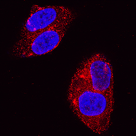
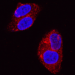
- Glypican 3 in HepG2 Human Cell Line. Glypican 3 was detected in immersion fixed HepG2 human hepatocellular carcinoma cell line using Mouse Anti-Human Glypican 3 Monoclonal Antibody (Catalog # MAB2119) at 3 µg/mL for 3 hours at room temperature. Cells were stained using the NorthernLights™ 557-conjugated Anti-Mouse IgG Secondary Antibody (red; Catalog # NL007) and counterstained with DAPI (blue). Specific staining was localized to cytoplasm and cell membranes. View our protocol for Fluorescent ICC Staining of Cells on Coverslips.