Explore
 Validate
 Learn
Learn Western blot
 Immunocytochemistry
Antibody data
- Antibody Data
- Antigen structure
- References [22]
- Comments [0]
- Validations
- Immunocytochemistry [5]
- Immunohistochemistry [1]
- Other assay [8]
Submit
Validation data
Reference
Comment
Report error
- Product number
- MA1-914 - Provider product page

- Provider
- Invitrogen Antibodies
- Product name
- PMCA4 ATPase Monoclonal Antibody (JA9)
- Antibody type
- Monoclonal
- Antigen
- Purifed from natural sources
- Description
- MA1-914 specifically detects plasma membrane calcium pump PMCA4 ATPase from human, bovine, porcine and rat tissues. MA1-914 has been successfully used in Western blot, immunohistochemistry and ELISA procedures. By Western blot, this antibody detects an ~129 kDa and an ~133 kDa protein representing PMCA4b and PMCA4a ATPase, respectively. The MA1-914 antigen is purified human erythrocyte PMCA ATPase. The epitope for this antibody has been mapped to amino acids 51-75 of PMCA4 ATPase. This sequence is common to both PMCA4a and 4b ATPase.
- Reactivity
- Human, Rat, Bovine, Porcine
- Host
- Mouse
- Isotype
- IgG
- Antibody clone number
- JA9
- Vial size
- 100 μg
- Concentration
- 1 mg/mL
- Storage
- -20°C, Avoid Freeze/Thaw Cycles
Submitted references Acute Genetic Ablation of Cardiac Sodium/Calcium Exchange in Adult Mice: Implications for Cardiomyocyte Calcium Regulation, Cardioprotection, and Arrhythmia.
Metabolic regulation of calcium pumps in pancreatic cancer: role of phosphofructokinase-fructose-bisphosphatase-3 (PFKFB3).
Plasma Membrane Ca(2+) ATPase Isoform 4 (PMCA4) Has an Important Role in Numerous Hallmarks of Pancreatic Cancer.
Molecular and Electrophysiological Analyses of ATP2B4 Gene Variants in Bilateral Adrenal Hyperaldosteronism.
Glutamate Deregulation in Ketamine-Induced Psychosis-A Potential Role of PSD95, NMDA Receptor and PMCA Interaction.
PMCA4 (ATP2B4) mutation in familial spastic paraplegia causes delay in intracellular calcium extrusion.
Vesicular transfer of membrane components to bovine epididymal spermatozoa.
Distinct regulation of cytoplasmic calcium signals and cell death pathways by different plasma membrane calcium ATPase isoforms in MDA-MB-231 breast cancer cells.
Plasma membrane calcium-ATPase isoform four distribution changes during corneal epithelial wound healing.
Muscarinic-induced recruitment of plasma membrane Ca2+-ATPase involves PSD-95/Dlg/Zo-1-mediated interactions.
Compartmentalized expression of three novel sarco/endoplasmic reticulum Ca2+ATPase 3 isoforms including the switch to ER stress, SERCA3f, in non-failing and failing human heart.
Caytaxin deficiency disrupts signaling pathways in cerebellar cortex.
Differential regulation of the apical plasma membrane Ca(2+) -ATPase by protein kinase A in parotid acinar cells.
Plasma membrane calcium pumps in mouse olfactory sensory neurons.
The plasma membrane Ca2+-ATPase isoform 4 is localized in lipid rafts of cerebellum synaptic plasma membranes.
Expression and immunolocalization of plasma membrane calcium ATPase isoforms in human corneal epithelium.
Expression of calcium transporters in the retina of the tiger salamander (Ambystoma tigrinum).
Regional distribution of Na,K-ATPase activity in porcine lens epithelium.
Regional distribution of Na,K-ATPase activity in porcine lens epithelium.
Plasma membrane Ca2+ pump in rat brain. Patterns of alternative splices seen by isoform-specific antibodies.
Detection of isoform 4 of the plasma membrane calcium pump in human tissues by using isoform-specific monoclonal antibodies.
Use of expression mutants and monoclonal antibodies to map the erythrocyte Ca2+ pump.
Lotteau S, Zhang R, Hazan A, Grabar C, Gonzalez D, Aynaszyan S, Philipson KD, Ottolia M, Goldhaber JI
Journal of the American Heart Association 2021 Sep 7;10(17):e019273
Journal of the American Heart Association 2021 Sep 7;10(17):e019273
Metabolic regulation of calcium pumps in pancreatic cancer: role of phosphofructokinase-fructose-bisphosphatase-3 (PFKFB3).
Richardson DA, Sritangos P, James AD, Sultan A, Bruce JIE
Cancer & metabolism 2020;8:2
Cancer & metabolism 2020;8:2
Plasma Membrane Ca(2+) ATPase Isoform 4 (PMCA4) Has an Important Role in Numerous Hallmarks of Pancreatic Cancer.
Sritangos P, Pena Alarcon E, James AD, Sultan A, Richardson DA, Bruce JIE
Cancers 2020 Jan 16;12(1)
Cancers 2020 Jan 16;12(1)
Molecular and Electrophysiological Analyses of ATP2B4 Gene Variants in Bilateral Adrenal Hyperaldosteronism.
Hattangady NG, Foster J, Lerario AM, Ponce-Balbuena D, Rege J, Monticone S, Rainey WE, Mulatero P, Else T
Hormones & cancer 2020 Feb;11(1):52-62
Hormones & cancer 2020 Feb;11(1):52-62
Glutamate Deregulation in Ketamine-Induced Psychosis-A Potential Role of PSD95, NMDA Receptor and PMCA Interaction.
Lisek M, Ferenc B, Studzian M, Pulaski L, Guo F, Zylinska L, Boczek T
Frontiers in cellular neuroscience 2017;11:181
Frontiers in cellular neuroscience 2017;11:181
PMCA4 (ATP2B4) mutation in familial spastic paraplegia causes delay in intracellular calcium extrusion.
Ho PW, Pang SY, Li M, Tse ZH, Kung MH, Sham PC, Ho SL
Brain and behavior 2015 Apr;5(4):e00321
Brain and behavior 2015 Apr;5(4):e00321
Vesicular transfer of membrane components to bovine epididymal spermatozoa.
Schwarz A, Wennemuth G, Post H, Brandenburger T, Aumüller G, Wilhelm B
Cell and tissue research 2013 Sep;353(3):549-61
Cell and tissue research 2013 Sep;353(3):549-61
Distinct regulation of cytoplasmic calcium signals and cell death pathways by different plasma membrane calcium ATPase isoforms in MDA-MB-231 breast cancer cells.
Curry MC, Luk NA, Kenny PA, Roberts-Thomson SJ, Monteith GR
The Journal of biological chemistry 2012 Aug 17;287(34):28598-608
The Journal of biological chemistry 2012 Aug 17;287(34):28598-608
Plasma membrane calcium-ATPase isoform four distribution changes during corneal epithelial wound healing.
Talarico EF Jr
Molecular vision 2010 Nov 2;16:2259-72
Molecular vision 2010 Nov 2;16:2259-72
Muscarinic-induced recruitment of plasma membrane Ca2+-ATPase involves PSD-95/Dlg/Zo-1-mediated interactions.
Kruger WA, Yun CC, Monteith GR, Poronnik P
The Journal of biological chemistry 2009 Jan 16;284(3):1820-30
The Journal of biological chemistry 2009 Jan 16;284(3):1820-30
Compartmentalized expression of three novel sarco/endoplasmic reticulum Ca2+ATPase 3 isoforms including the switch to ER stress, SERCA3f, in non-failing and failing human heart.
Dally S, Monceau V, Corvazier E, Bredoux R, Raies A, Bobe R, del Monte F, Enouf J
Cell calcium 2009 Feb;45(2):144-54
Cell calcium 2009 Feb;45(2):144-54
Caytaxin deficiency disrupts signaling pathways in cerebellar cortex.
Xiao J, Gong S, Ledoux MS
Neuroscience 2007 Jan 19;144(2):439-61
Neuroscience 2007 Jan 19;144(2):439-61
Differential regulation of the apical plasma membrane Ca(2+) -ATPase by protein kinase A in parotid acinar cells.
Baggaley E, McLarnon S, Demeter I, Varga G, Bruce JI
The Journal of biological chemistry 2007 Dec 28;282(52):37678-93
The Journal of biological chemistry 2007 Dec 28;282(52):37678-93
Plasma membrane calcium pumps in mouse olfactory sensory neurons.
Weeraratne SD, Valentine M, Cusick M, Delay R, Van Houten JL
Chemical senses 2006 Oct;31(8):725-30
Chemical senses 2006 Oct;31(8):725-30
The plasma membrane Ca2+-ATPase isoform 4 is localized in lipid rafts of cerebellum synaptic plasma membranes.
Sepúlveda MR, Berrocal-Carrillo M, Gasset M, Mata AM
The Journal of biological chemistry 2006 Jan 6;281(1):447-53
The Journal of biological chemistry 2006 Jan 6;281(1):447-53
Expression and immunolocalization of plasma membrane calcium ATPase isoforms in human corneal epithelium.
Talarico EF Jr, Kennedy BG, Marfurt CF, Loeffler KU, Mangini NJ
Molecular vision 2005 Mar 2;11:169-78
Molecular vision 2005 Mar 2;11:169-78
Expression of calcium transporters in the retina of the tiger salamander (Ambystoma tigrinum).
Krizaj D, Liu X, Copenhagen DR
The Journal of comparative neurology 2004 Aug 2;475(4):463-80
The Journal of comparative neurology 2004 Aug 2;475(4):463-80
Regional distribution of Na,K-ATPase activity in porcine lens epithelium.
Tamiya S, Dean WL, Paterson CA, Delamere NA
Investigative ophthalmology & visual science 2003 Oct;44(10):4395-9
Investigative ophthalmology & visual science 2003 Oct;44(10):4395-9
Regional distribution of Na,K-ATPase activity in porcine lens epithelium.
Tamiya S, Dean WL, Paterson CA, Delamere NA
Investigative ophthalmology & visual science 2003 Oct;44(10):4395-9
Investigative ophthalmology & visual science 2003 Oct;44(10):4395-9
Plasma membrane Ca2+ pump in rat brain. Patterns of alternative splices seen by isoform-specific antibodies.
Filoteo AG, Elwess NL, Enyedi A, Caride A, Aung HH, Penniston JT
The Journal of biological chemistry 1997 Sep 19;272(38):23741-7
The Journal of biological chemistry 1997 Sep 19;272(38):23741-7
Detection of isoform 4 of the plasma membrane calcium pump in human tissues by using isoform-specific monoclonal antibodies.
Caride AJ, Filoteo AG, Enyedi A, Verma AK, Penniston JT
The Biochemical journal 1996 May 15;316 ( Pt 1):353-9
The Biochemical journal 1996 May 15;316 ( Pt 1):353-9
Use of expression mutants and monoclonal antibodies to map the erythrocyte Ca2+ pump.
Adamo HP, Caride AJ, Penniston JT
The Journal of biological chemistry 1992 Jul 15;267(20):14244-9
The Journal of biological chemistry 1992 Jul 15;267(20):14244-9
No comments: Submit comment
Supportive validation
- Submitted by
- Invitrogen Antibodies (provider)
- Main image
- Experimental details
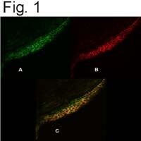
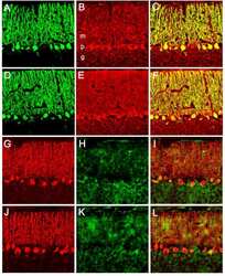
- Immunolocalization of PMCA4 and CaSRO in bovine corneal epithelium (bCE). (A) Immunolocalization of PMCA4 (green) using Product # MA1-914 on cell membranes of all cells in each layer of bCE. (B) Immunolocalization of CaSRO (red) using Product # MA1-934 on cell membranes of bCE mostly in wing- and squamous cell layers. (C) Merged image.

- Submitted by
- Invitrogen Antibodies (provider)
- Main image
- Experimental details
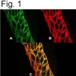
- Immunolocalization of PMCA4 and CaSRO in human corneal epithelium. (A) Immunolocalization of PMCA4 (green) using Product # MA1-914 on cell membranes of all cells in each layer of hCE. (B) Immunolocalization of CaSRO (red) using Product # MA1-934 on cell membranes of hCE. (C) Merged image.

- Submitted by
- Invitrogen Antibodies (provider)
- Main image

- Experimental details
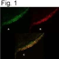
- Immunolocalization of PMCA4 and CaSRO in bovine corneal epithelium (bCE). (A) Immunolocalization of PMCA4 (green) using Product # MA1-914 on cell membranes of all cells in each layer of bCE. (B) Immunolocalization of CaSRO (red) using Product # MA1-934 on cell membranes of bCE mostly in wing- and squamous cell layers. (C) Merged image.

- Submitted by
- Invitrogen Antibodies (provider)
- Main image

- Experimental details
- Immunolocalization of PMCA4 and CaSRO in human corneal epithelium. (A) Immunolocalization of PMCA4 (green) using Product # MA1-914 on cell membranes of all cells in each layer of hCE. (B) Immunolocalization of CaSRO (red) using Product # MA1-934 on cell membranes of hCE. (C) Merged image.

- Submitted by
- Invitrogen Antibodies (provider)
- Main image

- Experimental details
- Immunolocalization of PMCA4 and CaSRO in bovine corneal epithelium (bCE). (A) Immunolocalization of PMCA4 (green) using Product # MA1-914 on cell membranes of all cells in each layer of bCE. (B) Immunolocalization of CaSRO (red) using Product # MA1-934 on cell membranes of bCE mostly in wing- and squamous cell layers. (C) Merged image.
Supportive validation
- Submitted by
- Invitrogen Antibodies (provider)
- Main image

- Experimental details
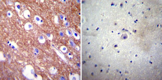
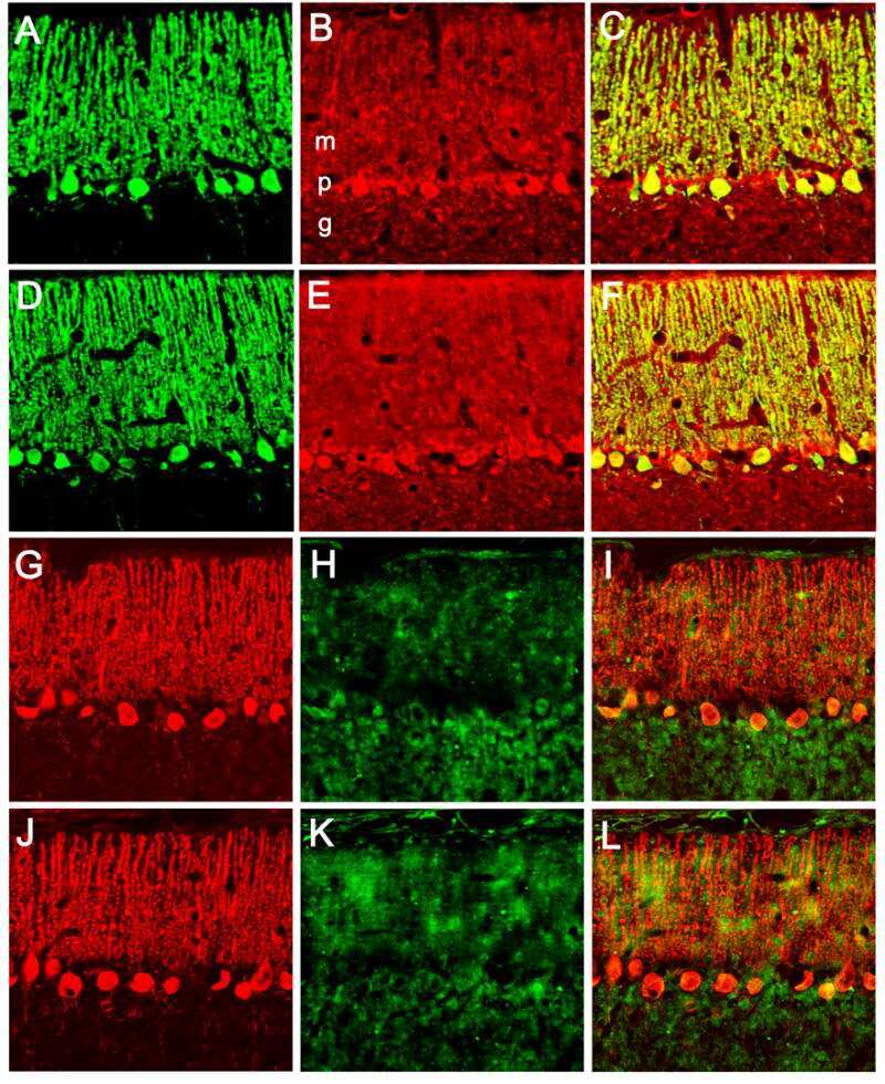
- Immunohistochemistry was performed on normal biopsies of deparaffinized Human brain tissue. To expose target proteins, heat induced antigen retrieval was performed using 10mM sodium citrate (pH6.0) buffer, microwaved for 8-15 minutes. Following antigen retrieval tissues were blocked in 3% BSA-PBS for 30 minutes at room temperature. Tissues were then probed at a dilution of 1:100 with a mouse monoclonal antibody recognizing PMCA4 ATPase (Product # MA1-914) or without primary antibody (negative control) overnight at 4°C in a humidified chamber. Tissues were washed extensively with PBST and endogenous peroxidase activity was quenched with a peroxidase suppressor. Detection was performed using a biotin-conjugated secondary antibody and SA-HRP, followed by colorimetric detection using DAB. Tissues were counterstained with hematoxylin and prepped for mounting.
Supportive validation
- Submitted by
- Invitrogen Antibodies (provider)
- Main image
- Experimental details
- NULL
- Submitted by
- Invitrogen Antibodies (provider)
- Main image
- Experimental details
- NULL
- Submitted by
- Invitrogen Antibodies (provider)
- Main image
- Experimental details
- NULL

- Submitted by
- Invitrogen Antibodies (provider)
- Main image
- Experimental details
- NULL
- Submitted by
- Invitrogen Antibodies (provider)
- Main image

- Experimental details
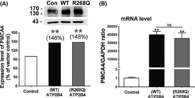
- Figure 1 (A) Western blot showing stable overexpression of either wild-type (WT) or R268Q mutant ATP2B4 protein at similar level in SH-SY5Y cells. (B) ATP2B4 mRNA levels in WT and mutant stably overexpressing cells were similar as shown by quantitative real-time PCR. ** represents statistical significance at p
- Submitted by
- Invitrogen Antibodies (provider)
- Main image
- Experimental details
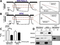
- Fig. 4 PFK15 causes PMCA inhibition in MIA PaCa-2 but not human pancreatic stellate cells: PMCA activity was measured in MIA PaCa-2 cells and HPSCs perfused under constant SERCA blockade (cyclopiazonic acid, CPA, 30 muM) and calcium-free conditions and 1 mM EGTA (black bar). Subsequent ER Ca 2+ depletion caused store-operated calcium entry when 20 mM Ca 2+ was introduced (white bar) calcium efflux was used as a measure of PMCA activity before and after 30 min incubation with or without 10 muM PFK15. Representative traces showing clearance experiments with PFK15 in MIA PaCa-2 ( a ) and HPSCs ( b ). The rate of clearance was calculated for the fastest 2 min of the first clearance phase ( R 1 ), the clearance rate from the same starting ratio was then calculated in the second clearance phase ( R 2 ). R 1 / R 2 was calculated for both MIA PaCa-2 and HPSC with and without PFK15 treatment ( c ). Membrane and cytosolic protein samples from MIA PaCa-2 cells were produced using a membrane protein extraction kit and PFKFB3 levels assessed by immunoblot. PMCA4 was used as a positive control for membrane samples and GAPDH was used as a positive control for cytoplasmic samples, a representative image is shown ( d ). Bars represent the mean +- SEM of 3-5 experiments. Kruskall-Wallis test; ** p < 0.005

- Submitted by
- Invitrogen Antibodies (provider)
- Main image
- Experimental details
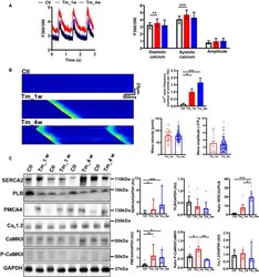
- Figure 6 Adaptations of calcium (Ca 2+ ) handling in tamoxifen-inducible sodium-Ca 2+ exchanger isoform 1 (NCX1) knockout (KO) mice. A , Representative traces (left) and summary data (right) of Ca 2+ transients recorded using Fura-2 acetoxymethyl ester in control mice (Ctl, black) and tamoxifen-inducible NCX1 KO mice at 1 week (Tm_1w, red) and 4 weeks (Tm_4w, blue) post-tamoxifen. B , Representative confocal line scans (left) and summary data (right) of Ca 2+ wave frequency, velocity (mum/s), and amplitude (DeltaF/F 0 ) recorded in isolated ventricular myocytes loaded with Fluo-4 acetoxymethyl ester from control mice (Ctl, N=66 cells from 8 mice) and mice 1 week (Tm_1w, N=47 cells from 4 mice) and 4 weeks (Tm_4w, N=62 cells from 6 mice) after tamoxifen injection. C , Representative Western blots (left) and summary data (right) from ventricular cardiomyocytes showing sarcoplasmic reticulum Ca 2+ ATPase 2 (SERCA2), phospholamban (PLB), plasma membrane calcium ATPase 4 (PMCA4), Ca v 1.2 (calcium channel voltage-dependent L type alpha 1C subunit), calcium/calmodulin-dependent protein kinase II (CaMKII), and phosphorylated CaMKII (P-CaMKII) expression. Mean data normalized to GAPDH. Data are expressed as mean+-SEM (1-way ANOVA) or median and interquartile range (Kruskal-Wallis). Data from control mice (Ctl, N=9) and mice 1 week (Tm_1w, N=6) and 4 weeks (Tm_4w, N=6 mice) after tamoxifen injection. * P
- Submitted by
- Invitrogen Antibodies (provider)
- Main image

- Experimental details
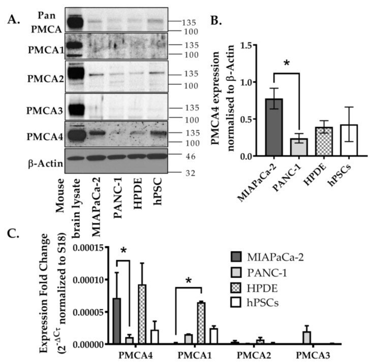
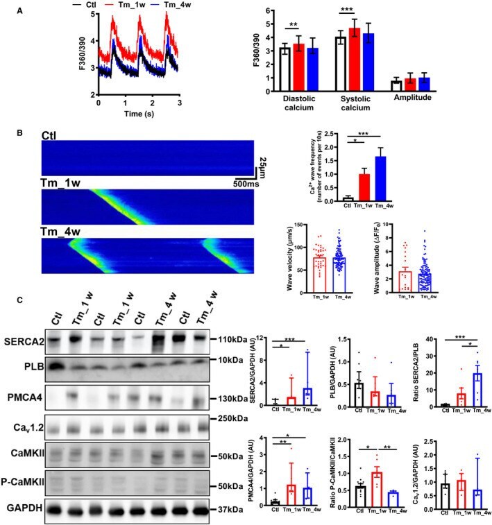
- Figure 2 Expression of PMCA isoforms in multiple pancreatic cell lines. ( A ) Representative Western immunoblot showing the relative protein expression of total/pan-PMCA and PMCA isoform 1-4 in pancreatic cancer (MIA PaCa-2 and PANC-1) and non-malignant pancreatic cells (human pancreatic ductal epithelial (HPDE) and human pancreatic stellate cells (hPSC)). Mouse brain lysate was used as a positive control for PMCA expressions and beta-Actin was used as a protein loading control. ( B ) PMCA4 protein expression in each cell line was quantified from Western blot bands and normalized to beta-Actin housekeeping protein. ( C ) The relative expressions of ATP2B1-4 (PMCA1-4 mRNA) in each cell line were quantified by RT-qPCR. Data are expressed as relative mRNA expression normalized to corresponding S18 rRNA controls (2 -DeltaCtau ). Statistical comparisons were made using the Kruskal-Wallis test with Dunn's multiple comparison test and two-way analysis of variance (ANOVA) with Dunnett's multiple comparison test. Data are expressed as mean +- SEM. (n = 4-5, 4 replicates per treatment condition). * represents statistical significance where p < 0.05.