 Explore
 Validate
 Learn
Learn Western blot
Antibody data
- Antibody Data
- Antigen structure
- References [0]
- Comments [0]
- Validations
- Western blot [1]
- Immunocytochemistry [1]
- Immunohistochemistry [1]
Submit
Validation data
Reference
Comment
Report error
- Product number
- AF5758 - Provider product page

- Provider
- R&D Systems
- Product name
- Human ING1 Antibody
- Antibody type
- Polyclonal
- Description
- Immunogen affinity purified. Detects human ING1 in direct ELISAs and Western blots. In direct ELISAs, approximately 5% cross-reactivity with recombinant human (rh) ING2 and less than 1% cross-reactivity with with rhING3, rhING4, and rhING5 is observed.
- Reactivity
- Human
- Host
- Goat
- Conjugate
- Unconjugated
- Antigen sequence
Q9UK53- Isotype
- IgG
- Vial size
- 100 ug
- Concentration
- LYOPH
- Storage
- Use a manual defrost freezer and avoid repeated freeze-thaw cycles. 12 months from date of receipt, -20 to -70 °C as supplied. 1 month, 2 to 8 °C under sterile conditions after reconstitution. 6 months, -20 to -70 °C under sterile conditions after reconstitution.
No comments: Submit comment
Supportive validation
- Submitted by
- R&D Systems (provider)
- Main image
- Experimental details
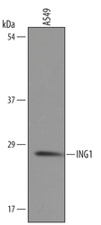
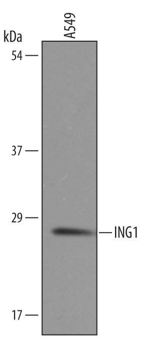
- Detection of Human ING1 by Western Blot. Western blot shows lysates of A549 human lung carcinoma cell line . PVDF membrane was probed with 1 µg/mL of Goat Anti-Human ING1 Antigen Affinity-purified Polyclonal Antibody (Catalog # AF5758) followed by HRP-conjugated Anti-Goat IgG Secondary Antibody (Catalog # HAF019). A specific band was detected for ING1 at approximately 28 kDa (as indicated). This experiment was conducted under reducing conditions and using Immunoblot Buffer Group 8.
Supportive validation
- Submitted by
- R&D Systems (provider)
- Main image
- Experimental details
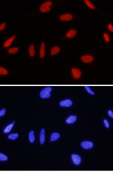
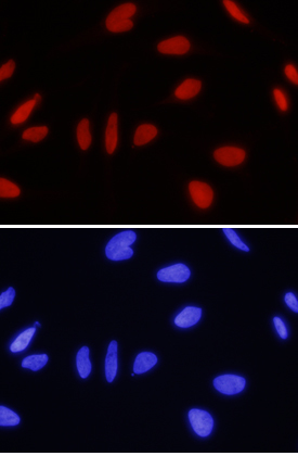
- ING1 in HeLa Human Cell Line. ING1 was detected in immersion fixed HeLa human cervical epithelial carcinoma cell line using 10 µg/mL Goat Anti-Human ING1 Antigen Affinity-purified Polyclonal Antibody (Catalog # AF5758) for 3 hours at room temperature. Cells were stained with the NorthernLights™ 557-conjugated Anti-Goat IgG Secondary Antibody (red, upper panel; Catalog # NL001) and counterstained with DAPI (blue, lower panel). View our protocol for Chromogenic IHC Staining of Paraffin-embedded Tissue Sections.
Supportive validation
- Submitted by
- R&D Systems (provider)
- Main image
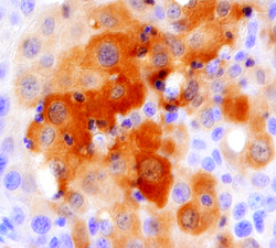
- Experimental details
- ING1 in Human Lung Cancer Tissue. ING1 was detected in immersion fixed paraffin-embedded sections of human lung cancer tissue using 5 µg/mL Goat Anti-Human ING1 Antigen Affinity-purified Polyclonal Antibody (Catalog # AF5758) overnight at 4 °C. Tissue was stained with the Anti-Goat HRP-DAB Cell & Tissue Staining Kit (brown; Catalog # CTS008) and counterstained with hematoxylin (blue). View our protocol for Fluorescent ICC Staining of Cells on Coverslips.
