 Explore
 Validate
 Learn
Learn Western blot
 ELISA
ELISA Immunocytochemistry
Antibody data
- Antibody Data
- Antigen structure
- References [1]
- Comments [0]
- Validations
- Immunocytochemistry [2]
- Immunohistochemistry [1]
- Other assay [2]
Submit
Validation data
Reference
Comment
Report error
- Product number
- PA5-97822 - Provider product page

- Provider
- Invitrogen Antibodies
- Product name
- HBG2 Polyclonal Antibody
- Antibody type
- Polyclonal
- Antigen
- Recombinant full-length protein
- Reactivity
- Human, Mouse, Rat
- Host
- Rabbit
- Isotype
- IgG
- Vial size
- 100 μg
- Concentration
- 1 mg/mL
- Storage
- -20°C or -80°C if preferred
Submitted references EPCR-PAR1 biased signaling regulates perfusion recovery and neovascularization in peripheral ischemia.
Bochenek ML, Gogiraju R, Großmann S, Krug J, Orth J, Reyda S, Georgiadis GS, Spronk HM, Konstantinides S, Münzel T, Griffin JH, Wild P, Espinola-Klein C, Ruf W, Schäfer K
JCI insight 2022 Jul 22;7(14)
JCI insight 2022 Jul 22;7(14)
No comments: Submit comment
Supportive validation
- Submitted by
- Invitrogen Antibodies (provider)
- Main image

- Experimental details
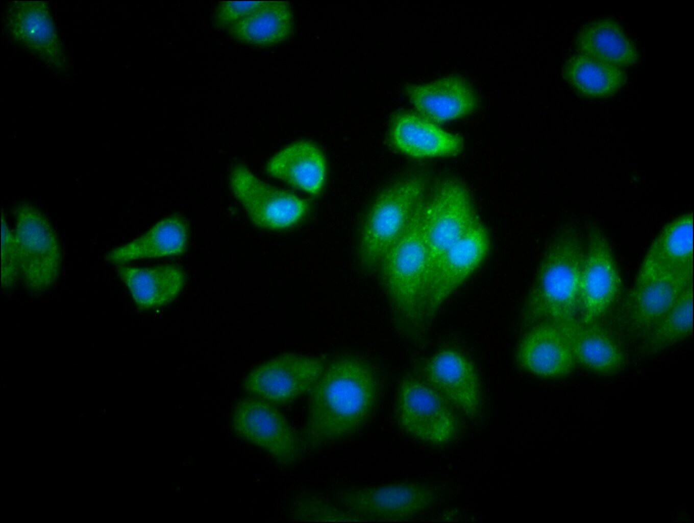
- Immunofluorescent analysis of HBG2 in HepG2 cells using a HBG2 polyclonal antibody (Product # PA5-97822) at a dilution of 1:100. The cells were fixed in 4% formaldehyde, permeabilized using 0.2% Triton X-100 and blocked in 10% normal Goat Serum. The cells were then incubated with the antibody overnight at 4°C. The secondary antibody was Alexa Fluor 488-congugated Goat Anti-Rabbit IgG(H+L). Cells were counter-stained with DAPI.

- Submitted by
- Invitrogen Antibodies (provider)
- Main image

- Experimental details
- Immunofluorescent analysis of HBG2 in HepG2 cells using a HBG2 polyclonal antibody (Product # PA5-97822) at a dilution of 1:100. The cells were fixed in 4% formaldehyde, permeabilized using 0.2% Triton X-100 and blocked in 10% normal Goat Serum. The cells were then incubated with the antibody overnight at 4°C. The secondary antibody was Alexa Fluor 488-congugated Goat Anti-Rabbit IgG(H+L). Cells were counter-stained with DAPI.
Supportive validation
- Submitted by
- Invitrogen Antibodies (provider)
- Main image
- Experimental details
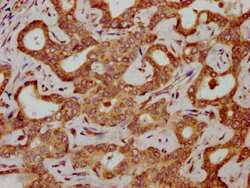
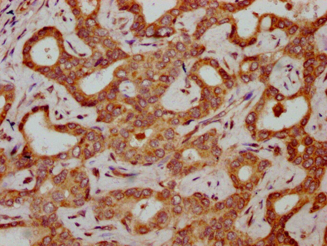
- Immunohistochemical analysis of HBG2 in paraffin embedded human liver cancer using a HBG2 polyclonal antibody (Product # PA5-97822) at a dilution of 1:300. After dewaxing and hydration, antigen retrieval was mediated by high pressure in a citrate buffer (pH 6.0). Section was blocked with 10% normal goat serum 30min at RT. Then primary antibody (1% BSA) was incubated at 4°C overnight. The primary is detected by a biotinylated secondary antibody and visualized using an HRP conjugated SP system.
Supportive validation
- Submitted by
- Invitrogen Antibodies (provider)
- Main image
- Experimental details
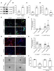
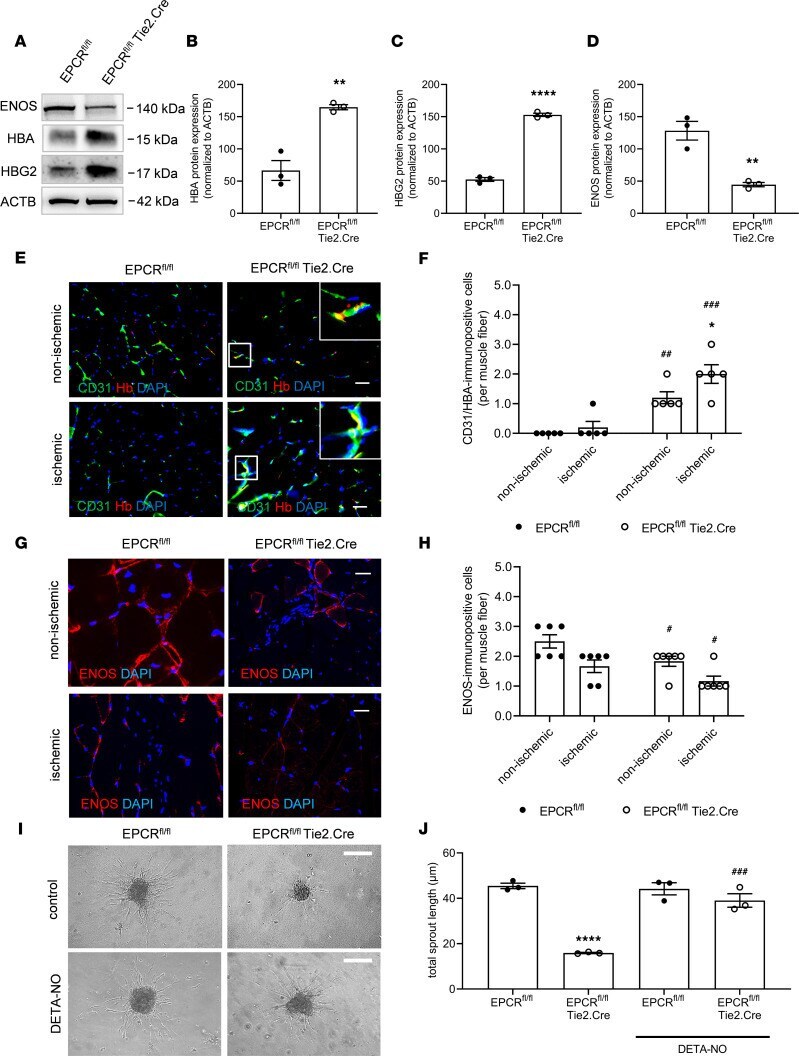
- Figure 4 EPCR-dependent hemoglobin expression and its effects on nitric oxide bioavailability and function. ( A - D ) Representative western blot membranes and their quantification showing HBA, HBG2 (methemoglobin), and eNOS protein levels in murine primary endothelial cells isolated from lungs of EPCR fl/fl Tie2.Cre mice and EPCR fl/fl littermate controls; n = 3 biological replicates. ** P < 0.01 and **** P < 0.0001 versus EPCR fl/fl mice; unpaired Student's t test. ( E and F ) Representative confocal images and the results of the quantitative analysis after visualization of HBA (red) expression in CD31 + endothelial cells (green) on cryopreserved cross sections isolated at day 28 after ischemia; n = 5 biological replicates. DAPI + cell nuclei appear blue. Scale bars: 10 mum. Note that myoglobin expression was not detected in endothelial cells in EPCR fl/fl Tie2.Cre mice and their littermate controls. ( G and H ) Representative confocal microscopy images and quantification of cryopreserved cross sections of nonischemic and ischemic hindlimbs of EPCR fl/fl Tie2.Cre mice and EPCR fl/fl littermate controls stained using antibodies against eNOS (red). DAPI + cell nuclei appear blue. Scale bars: 10 mum; n = 3 biological replicates per 2 experimental repeats; # P < 0.05 versus EPCR fl/fl control littermate mice using 2-way ANOVA, Sidak's multiple-comparison test. ( I and J ) Representative microscopy brightfield images and quantification of total sprout length in control and DETA-
- Submitted by
- Invitrogen Antibodies (provider)
- Main image
- Experimental details
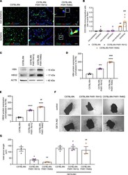
- Endothelial hemoglobin expression and NO-dependent angiogenesis in PAR1 mutant mice. ( A and B ) Representative confocal immunofluorescence microscopy images and quantitative analysis after visualization of HBA (red) expression in CD31 + endothelial cells (green) on cryopreserved cross sections isolated from mice at day 28 after ischemia. DAPI + cell nuclei appear blue. Scale bars: 10 mum. Two-way ANOVA, Sidak's multiple comparison; n = 5 biological replicates. * P < 0.05 versus nonischemic hindlimb; ## P < 0.01 and ### P < 0.001 versus C57BL/6N control mice. ( C - E ) Representative Western blots and quantification of HBA and HBG2 (methemoglobin) protein levels in primary lung endothelial cells isolated from C57BL/6N, C57BL/6N PAR1 R41Q, and C57BL/6N PAR1 R46Q mutant mice; n = 3 biological replicates. Samples were run on the same blot. Nonadjacent lanes are indicated by an empty space between them. One-way ANOVA, Sidak's multiple comparison. *** P < 0.001 and **** P < 0.0001 versus C57BL/6N; ## P < 0.01 and #### P < 0.0001 versus C57BL/6N PAR1 R41Q. Angiogenic sprout formation from aortic rings of C57BL/6N, C57BL/6N PAR1 R41Q, and C57BL/6N PAR1 R46Q mutant mice with and without DETA-NO (100 muM). ( F and G ) Representative findings and quantitative analysis are shown; n = 3 biological replicates. Scale bars: 100 mum. * P < 0.05 and ** P < 0.01 versus C57BL/6N; # P < 0.05 and ## P < 0.01 for DETA-NO versus untreated aortic rings of the same group. One-way ANOVA, Sidak's multi
