 Explore
 Validate
 Learn
Learn Immunocytochemistry
Antibody data
- Antibody Data
- Antigen structure
- References [1]
- Comments [0]
- Validations
- Immunocytochemistry [1]
- Flow cytometry [1]
Submit
Validation data
Reference
Comment
Report error
- Product number
- 11-1169-42 - Provider product page

- Provider
- Invitrogen Antibodies
- Product name
- CD116 Monoclonal Antibody (4H1), FITC, eBioscience™
- Antibody type
- Monoclonal
- Antigen
- Other
- Description
- Description: The 4H1 monoclonal antibody reacts with the human CD116 molecule, the alpha subunit of GM-CSF receptor. The alpha subunit associates with the common beta chain (CD131) to form the high affinity receptor for GM-CSF. The GM-CSFR alpha chain is expressed by granulocytes, monocytes, endothelial cells, fibroblasts and some tumor cells. Applications Reported: 4H1 has been reported for use in flow cytometric analysis. Applications Tested: This 4H1 antibody has been pre-titrated and tested by flow cytometric analysis of normal human peripheral blood cells. This can be used at 5 µL (1 µg) per test. A test is defined as the amount (µg) of antibody that will stain a cell sample in a final volume of 100 µL. Cell number should be determined empirically but can range from 10^5 to 10^8 cells/test. Excitation: 488 nm; Emission: 520 nm; Laser: Blue Laser. Filtration: 0.2 µm post-manufacturing filtered.
- Reactivity
- Human
- Host
- Mouse
- Conjugate
- Green dye
- Isotype
- IgG
- Antibody clone number
- 4H1
- Vial size
- 100 Tests
- Concentration
- 5 μL/Test
- Storage
- 4°C, store in dark, DO NOT FREEZE!
Submitted references Interaction between Hck and HIV-1 Nef negatively regulates cell surface expression of M-CSF receptor.
Hiyoshi M, Suzu S, Yoshidomi Y, Hassan R, Harada H, Sakashita N, Akari H, Motoyoshi K, Okada S
Blood 2008 Jan 1;111(1):243-50
Blood 2008 Jan 1;111(1):243-50
No comments: Submit comment
Supportive validation
- Submitted by
- Invitrogen Antibodies (provider)
- Main image
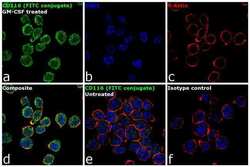
- Experimental details
- Immunofluorescence analysis of CD116 was performed using 70% confluent log phase THP-1 cells treated with 10 ng of GM-CSF for 15 minutes. The cells were fixed with 4% paraformaldehyde for 10 minutes, permeabilized with 0.1% Triton™ X-100 for 10 minutes, and blocked with 1% BSA for 1 hour at room temperature. The cells were labeled with CD116, FITC, Mouse Monoclonal antibody (Product # 11-1169-42) at 1:100 dilution in 0.1% BSA and incubated at 4 degree Celsius overnight (Panel a: green). Nuclei (Panel b: blue) were stained with SlowFade® Gold Antifade Mountant with DAPI (Product # S36938). F-actin (Panel c: red) was stained with Rhodamine Phalloidin (Product # R415, 1:300). Panel d represents the merged image showing membrane localization. Panel e shows untreated cells with less membrane signal. Panel f represents the isotype control. The images were captured at 60X magnification.
Supportive validation
- Submitted by
- Invitrogen Antibodies (provider)
- Main image
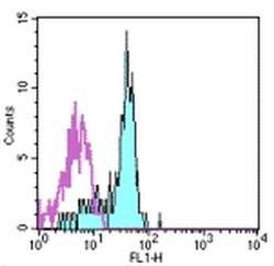
- Experimental details
- Staining of normal human peripheral blood cells with Mouse IgG1 kappa Isotype Control FITC (Product # 11-4714-42) (open histogram) or Anti-Human CD116 FITC (filled histogram). Cells in the monocyte gate were used for analysis.
