Explore
 Validate
 Learn
Learn Western blot
Antibody data
- Antibody Data
- Antigen structure
- References [1]
- Comments [0]
- Validations
- Western blot [1]
- Immunohistochemistry [1]
Submit
Validation data
Reference
Comment
Report error
- Product number
- MAB7940 - Provider product page

- Provider
- R&D Systems
- Product name
- Human GFER/ALR Antibody
- Antibody type
- Monoclonal
- Description
- Protein A or G purified from hybridoma culture supernatant. Detects human GFER/ALR in ELISAs.
- Reactivity
- Human
- Host
- Mouse
- Conjugate
- Unconjugated
- Antigen sequence
P55789- Isotype
- IgG
- Antibody clone number
- 829812
- Vial size
- 100 ug
- Concentration
- LYOPH
- Storage
- Use a manual defrost freezer and avoid repeated freeze-thaw cycles. 12 months from date of receipt, -20 to -70 °C as supplied. 1 month, 2 to 8 °C under sterile conditions after reconstitution. 6 months, -20 to -70 °C under sterile conditions after reconstitution.
Submitted references ERV1 Overexpression in Myeloid Cells Protects against High Fat Diet Induced Obesity and Glucose Intolerance.
Sima C, Montero E, Nguyen D, Freire M, Norris P, Serhan CN, Van Dyke TE
Scientific reports 2017 Oct 9;7(1):12848
Scientific reports 2017 Oct 9;7(1):12848
No comments: Submit comment
Supportive validation
- Submitted by
- R&D Systems (provider)
- Main image
- Experimental details
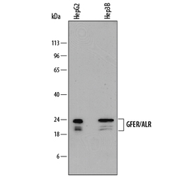
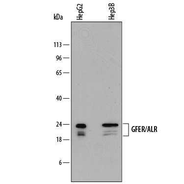
- Detection of Human GFER/ALR by Western Blot. Western blot shows lysates of HepG2 human hepatocellular carcinoma cell line and Hep3B human hepatocellular carcinoma cell line. PVDF membrane was probed with 2 µg/mL of Mouse Anti-Human GFER/ALR Monoclonal Antibody (Catalog # MAB7940) followed by HRP-conjugated Anti-Mouse IgG Secondary Antibody (Catalog # HAF018). Specific bands were detected for GFER/ALR at approximately 20-24 kDa (as indicated). This experiment was conducted under reducing conditions and using Immunoblot Buffer Group 1.
Supportive validation
- Submitted by
- R&D Systems (provider)
- Main image

- Experimental details
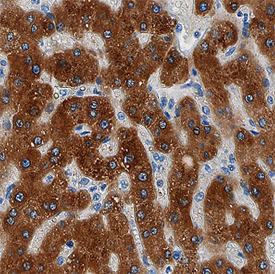
- GFER/ALR in Human Liver. GFER/ALR was detected in immersion fixed paraffin-embedded sections of human liver using Mouse Anti-Human GFER/ALR Monoclonal Antibody (Catalog # MAB7940) at 15 µg/mL overnight at 4 °C. Before incubation with the primary antibody, tissue was subjected to heat-induced epitope retrieval using Antigen Retrieval Reagent-Basic (Catalog # CTS013). Tissue was stained using the Anti-Mouse HRP-DAB Cell & Tissue Staining Kit (brown; Catalog # CTS002) and counter-stained with hematoxylin (blue). Specific staining was localized to cytoplasm of hepatocytes. View our protocol for Chromogenic IHC Staining of Paraffin-embedded Tissue Sections.