Explore
 Validate
 Learn
Learn Flow cytometry
Antibody data
- Antibody Data
- Antigen structure
- References [12]
- Comments [0]
- Validations
- Flow cytometry [1]
- Other assay [3]
Submit
Validation data
Reference
Comment
Report error
- Product number
- 12-9957-41 - Provider product page

- Provider
- Invitrogen Antibodies
- Product name
- HLA-G Monoclonal Antibody (87G), PE, eBioscience™
- Antibody type
- Monoclonal
- Antigen
- Other
- Description
- Description: The monoclonal antibody 87G recognizes human HLA-G, a member of the Human Leukocyte Antigen family but as part of the nonclassical MHC type involved in inhibiting immune reponses. HLA-G has seven reported isoforms. The antibody 87G recognizes both HLA-G1 and the solube HLA-G5. Expression of HLA-G is found primarily in fetal trophoblast cells as they invade the maternal decidua thereby protecting the fetus from the maternal immune system. Like the highly mitotic trophoblast, abundant HLA-G protein expression has been identified in some tumors, including melanoma, breast carcinoma and renal carcinoma as well as CLL, AML and B-CLL. Some expression has also been found in pancreatic islets, erythroid and endothelial progenitors and the adult thymic medulla.HLA-G+ CD4 or CD8 cells have been identified in normal human peripheral blood and are thought to act as regulatory cells in that they are hypoproliferative with a unique cytokine profile differing from Tregs. The receptors for HLA-G are CD85j/ILT2, CD85d/ILT4, and CD158. Recent studies have shown a role for HLA-G in tolerance and maintenance of transplanted organs.
- Conjugate
- Yellow dye
- Antibody clone number
- 87G
- Concentration
- 5 µL/Test
Submitted references Placental endovascular extravillous trophoblasts (enEVTs) educate maternal T-cell differentiation along the maternal-placental circulation.
hESC-derived immune suppressive dendritic cells induce immune tolerance of parental hESC-derived allografts.
Feasibility of placenta-derived mesenchymal stem cells as a tool for studying pregnancy-related disorders.
ADAM12-directed ectodomain shedding of E-cadherin potentiates trophoblast fusion.
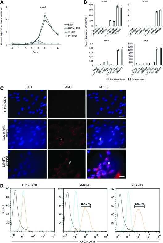
L3MBTL1 deficiency directs the differentiation of human embryonic stem cells toward trophectoderm.
Beyond the increasing complexity of the immunomodulatory HLA-G molecule.
HLA-G in cancer: a way to turn off the immune system.
Protein expression and peptide binding suggest unique and interacting functional roles for HLA-E, F, and G in maternal-placental immune recognition.
HLA-G, -E, -F preworkshop: tools and protocols for analysis of non-classical class I genes transcription and protein expression.
Modulation of HLA-G antigens expression in myelomonocytic cells.
Expression of HLA-G in human mononuclear phagocytes and selective induction by IFN-gamma.
Homotypic aggregation of human cell lines by HLA class II-, class Ia- and HLA-G-specific monoclonal antibodies.
Ma Y, Yang Q, Fan M, Zhang L, Gu Y, Jia W, Li Z, Wang F, Li YX, Wang J, Li R, Shao X, Wang YL
Cell proliferation 2020 May;53(5):e12802
Cell proliferation 2020 May;53(5):e12802
hESC-derived immune suppressive dendritic cells induce immune tolerance of parental hESC-derived allografts.
Todorova D, Zhang Y, Chen Q, Liu J, He J, Fu X, Xu Y
EBioMedicine 2020 Dec;62:103120
EBioMedicine 2020 Dec;62:103120
Feasibility of placenta-derived mesenchymal stem cells as a tool for studying pregnancy-related disorders.
Fuchi N, Miura K, Doi H, Li TS, Masuzaki H
Scientific reports 2017 Apr 12;7:46220
Scientific reports 2017 Apr 12;7:46220
ADAM12-directed ectodomain shedding of E-cadherin potentiates trophoblast fusion.
Aghababaei M, Hogg K, Perdu S, Robinson WP, Beristain AG
Cell death and differentiation 2015 Dec;22(12):1970-84
Cell death and differentiation 2015 Dec;22(12):1970-84
L3MBTL1 deficiency directs the differentiation of human embryonic stem cells toward trophectoderm.
Hoya-Arias R, Tomishima M, Perna F, Voza F, Nimer SD
Stem cells and development 2011 Nov;20(11):1889-900
Stem cells and development 2011 Nov;20(11):1889-900
Beyond the increasing complexity of the immunomodulatory HLA-G molecule.
Carosella ED, Favier B, Rouas-Freiss N, Moreau P, Lemaoult J
Blood 2008 May 15;111(10):4862-70
Blood 2008 May 15;111(10):4862-70
HLA-G in cancer: a way to turn off the immune system.
Rouas-Freiss N, Moreau P, Menier C, Carosella ED
Seminars in cancer biology 2003 Oct;13(5):325-36
Seminars in cancer biology 2003 Oct;13(5):325-36
Protein expression and peptide binding suggest unique and interacting functional roles for HLA-E, F, and G in maternal-placental immune recognition.
Ishitani A, Sageshima N, Lee N, Dorofeeva N, Hatake K, Marquardt H, Geraghty DE
Journal of immunology (Baltimore, Md. : 1950) 2003 Aug 1;171(3):1376-84
Journal of immunology (Baltimore, Md. : 1950) 2003 Aug 1;171(3):1376-84
HLA-G, -E, -F preworkshop: tools and protocols for analysis of non-classical class I genes transcription and protein expression.
Paul P, Rouas-Freiss N, Moreau P, Cabestre FA, Menier C, Khalil-Daher I, Pangault C, Onno M, Fauchet R, Martinez-Laso J, Morales P, Villena AA, Giacomini P, Natali PG, Frumento G, Ferrara GB, McMaster M, Fisher S, Schust D, Ferrone S, Dausset J, Geraghty D, Carosella ED
Human immunology 2000 Nov;61(11):1177-95
Human immunology 2000 Nov;61(11):1177-95
Modulation of HLA-G antigens expression in myelomonocytic cells.
Onno M, Le Friec G, Pangault C, Amiot L, Guilloux V, Drénou B, Caulet-Maugendre S, André P, Fauchet R
Human immunology 2000 Nov;61(11):1086-94
Human immunology 2000 Nov;61(11):1086-94
Expression of HLA-G in human mononuclear phagocytes and selective induction by IFN-gamma.
Yang Y, Chu W, Geraghty DE, Hunt JS
Journal of immunology (Baltimore, Md. : 1950) 1996 Jun 1;156(11):4224-31
Journal of immunology (Baltimore, Md. : 1950) 1996 Jun 1;156(11):4224-31
Homotypic aggregation of human cell lines by HLA class II-, class Ia- and HLA-G-specific monoclonal antibodies.
Odum N, Ledbetter JA, Martin P, Geraghty D, Tsu T, Hansen JA, Gladstone P
European journal of immunology 1991 Sep;21(9):2121-31
European journal of immunology 1991 Sep;21(9):2121-31
No comments: Submit comment
Supportive validation
- Submitted by
- Invitrogen Antibodies (provider)
- Main image
- Experimental details
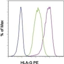
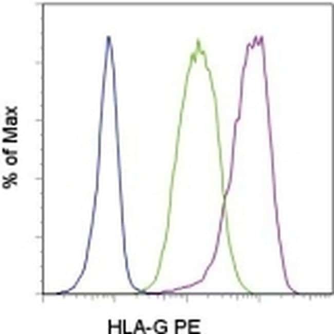
- Staining of GM-CSF and IFN gamma-stimulated U937 cells with Mouse IgG2a kappa Isotype Control PE (Product # 12-4724-81) (blue histogram) or Anti-Human HLA-G PE (purple histogram). Unstimulated U937 cells stained with Anti-Human HLA-G PE are shown in the green histogram. Total viable cells were used for analysis.
- Conjugate
- Yellow dye
Supportive validation
- Submitted by
- Invitrogen Antibodies (provider)
- Main image
- Experimental details
- NULL
- Conjugate
- Yellow dye
- Submitted by
- Invitrogen Antibodies (provider)
- Main image
- Experimental details
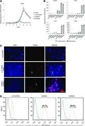
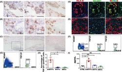
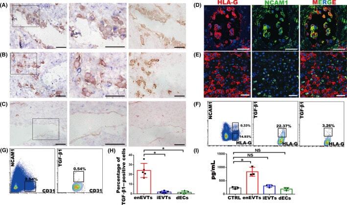
- FIGURE 2 Identification of TGF-beta1-producing cells at the maternal-foetal interface in normal pregnancy at gestational weeks 7-9. A-C, In situ hybridization of TGF-beta1 (blue) and immunohistochemistry staining of HLA-G (yellow in A and B) or CD31 (yellow in C) in normal pregnant decidua. Middle panels are enlargement of the rectangular areas in left panels, showing the enEVTs in remodelled SPA (A), iEVTs in the area nearby the remodelled SPA (B) and dECs in unremodelled SPA (C). Right panels are negative control of in situ hybridization, with the immunohistochemistry signals of HLA-G (A, B) or CD31 (C). D, E, Immunofluorescence of HLA-G (red) and NCAM1 (green) in remodelled SPA (D) and the area nearby remodelled SPA (E). F, Flow cytometry of TGF-beta1 expression in EVTs of normal pregnancy. Left panel, FACS isolation of enEVTs and iEVTs with antibodies against HLA-G and NCAM1. Middle panel, flow cytometry of TGF-beta1-positive enEVTs that are gated from the left panel as HLA-G + NCAM1 + . Right panel: flow cytometry analysis of TGF-beta1-positive iEVTs that were gated from the left panel as HLA-G + NCAM1 - . G, FACS isolation of CD31 + NCAM1 - dECs in normal pregnancy (left panel), and flow cytometry of TGF-beta1-positive dECs gated from the left panel as CD31 + NCAM1 - (right panel). H, The statistical analysis of TGF-beta1-positive primary cells based on the results from 5 normal pregnant cases. I, ELISA for TGF-beta1 in supernatants of the FACS-sorted enEVTs, iEVTs and
- Conjugate
- Yellow dye
- Submitted by
- Invitrogen Antibodies (provider)
- Main image

- Experimental details
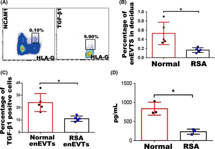
- FIGURE 3 The number of enEVTs and TGF-beta1-producing enEVTs in RSA pregnancy. A, Flow cytometry of enEVTs with antibodies against HLA-G and NCAM1 in RSA pregnancy (left panel). Flow cytometry of TGF-beta1-positive enEVTs that are gated from the left panel as HLA-G + NCAM1 + in RSA pregnancy (right panel). B, The statistical analysis of the number of enEVTs in normal (n = 5) and RSA (n = 5) pregnancy. C, The statistical analysis of the proportion of TGF-beta1-producing enEVTs in normal (n = 5) and RSA (n = 5) pregnancy. D, ELISA for TGF-beta1 in supernatants of the normal enEVTs (n = 3) and RSA enEVTs (n = 3). Data are presented as mean +- SD, and comparison between groups was performed with Student's t test. *, P < .05
- Conjugate
- Yellow dye