Explore
 Validate
 Learn
Learn Western blot
 Immunocytochemistry
Antibody data
- Antibody Data
- Antigen structure
- References [3]
- Comments [0]
- Validations
- Western blot [1]
- Immunohistochemistry [2]
Submit
Validation data
Reference
Comment
Report error
- Product number
- NB500-302 - Provider product page

- Provider
- Novus Biologicals
- Proper citation
- Novus Cat#NB500-302, RRID:AB_10003419
- Product name
- Mouse Monoclonal HLA G Antibody
- Antibody type
- Monoclonal
- Description
- Protein A purified. The antibody MEM-G/1 reacts with denaturated HLA-G heavy chain. HLA-G belongs to the MHC Class I molecules (MHC Class Ib
- Reactivity
- Human
- Host
- Mouse
- Isotype
- IgG
- Vial size
- 0.1 mg
- Concentration
- 1 mg/ml
- Storage
- Store at 4C. Do not freeze.
Submitted references Peptide hormone ELABELA enhances extravillous trophoblast differentiation, but placenta is not the major source of circulating ELABELA in pregnancy.
Notch1 controls development of the extravillous trophoblast lineage in the human placenta.
Notch signaling plays a critical role in motility and differentiation of human first-trimester cytotrophoblasts.
Georgiadou D, Boussata S, Ranzijn WHM, Root LEA, Hillenius S, Bij de Weg JM, Abheiden CNH, de Boer MA, de Vries JIP, Vrijkotte TGM, Lambalk CB, Kuijper EAM, Afink GB, van Dijk M
Scientific reports 2019 Dec 13;9(1):19077
Scientific reports 2019 Dec 13;9(1):19077
Notch1 controls development of the extravillous trophoblast lineage in the human placenta.
Haider S, Meinhardt G, Saleh L, Fiala C, Pollheimer J, Knöfler M
Proceedings of the National Academy of Sciences of the United States of America 2016 Nov 29;113(48):E7710-E7719
Proceedings of the National Academy of Sciences of the United States of America 2016 Nov 29;113(48):E7710-E7719
Notch signaling plays a critical role in motility and differentiation of human first-trimester cytotrophoblasts.
Haider S, Meinhardt G, Velicky P, Otti GR, Whitley G, Fiala C, Pollheimer J, Knöfler M
Endocrinology 2014 Jan;155(1):263-74
Endocrinology 2014 Jan;155(1):263-74
No comments: Submit comment
Supportive validation
- Submitted by
- Novus Biologicals (provider)
- Main image

- Experimental details
- Western Blot: HLA G Antibody (MEM-G/1) [NB500-302] - Using the Biotin direct conjugate analysis (reducing conditions) of HLA-G1 in HLA-G1 transfectants using the antibody MEM-G/1 biotin.

Supportive validation
- Submitted by
- Novus Biologicals (provider)
- Main image

- Experimental details
- Immunohistochemistry: HLA G Antibody (MEM-G/1) [NB500-302] - Fig. 1B - first-trimester placenta (paraffin-embedded sections)

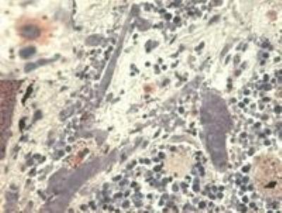
- Submitted by
- Novus Biologicals (provider)
- Main image

- Experimental details
- Immunohistochemistry: HLA G Antibody (MEM-G/1) [NB500-302] - Fig. 1. Immunohistochemistry staining with anti-human HLA-G (MEM-G/1). Fig. 1A - pulmonary disseases (paraffin-embedded sections) The antibody MEM-G/1 stains infiltrating macrophages in pulmonary diseases. In the top left corner see the detail of macrophage.