Explore
 Validate
 Learn
Learn Western blot
Antibody data
- Antibody Data
- Antigen structure
- References [3]
- Comments [0]
- Validations
- Western blot [2]
- Immunohistochemistry [2]
- Other assay [5]
Submit
Validation data
Reference
Comment
Report error
- Product number
- MA1-19219 - Provider product page

- Provider
- Invitrogen Antibodies
- Product name
- HLA-G Monoclonal Antibody (MEM-G/1)
- Antibody type
- Monoclonal
- Antigen
- Recombinant full-length protein
- Description
- This antibody reacts with an extracellular epitope of denaturated HLA-G heavy chain. HLA-G belongs to the MHC Class I molecules (MHC Class Ib; nonclassical) and it is expressed on the surface of trophoblast cells. Immunohistochemistry (Paraffin): Incubation: 1 h at RT.
- Reactivity
- Human
- Host
- Mouse
- Isotype
- IgG
- Antibody clone number
- MEM-G/1
- Vial size
- 100 μg
- Concentration
- 1 mg/mL
- Storage
- 4°C, do not freeze
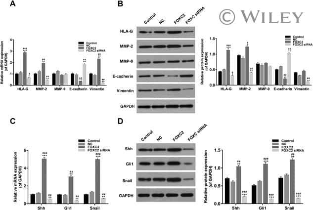
Submitted references Forkhead box C2 promotes the invasion ability of human trophoblast cells through Hedgehog (Hh) signaling pathway.
Differential expression of β-catenin and Dickkopf-1 in the third trimester placentas from normal and preeclamptic pregnancies: a comparative study.
Association of Wnt2 and sFRP4 expression in the third trimester placenta in women with severe preeclampsia.
Zhang Y, Zhang Y
Cell biology international 2018 Jul;42(7):859-866
Cell biology international 2018 Jul;42(7):859-866
Differential expression of β-catenin and Dickkopf-1 in the third trimester placentas from normal and preeclamptic pregnancies: a comparative study.
Zhang Z, Li H, Zhang L, Jia L, Wang P
Reproductive biology and endocrinology : RB&E 2013 Mar 4;11:17
Reproductive biology and endocrinology : RB&E 2013 Mar 4;11:17
Association of Wnt2 and sFRP4 expression in the third trimester placenta in women with severe preeclampsia.
Zhang Z, Zhang L, Zhang L, Jia L, Wang P, Gao Y
Reproductive sciences (Thousand Oaks, Calif.) 2013 Aug;20(8):981-9
Reproductive sciences (Thousand Oaks, Calif.) 2013 Aug;20(8):981-9
No comments: Submit comment
Supportive validation
- Submitted by
- Invitrogen Antibodies (provider)
- Main image
- Experimental details
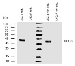
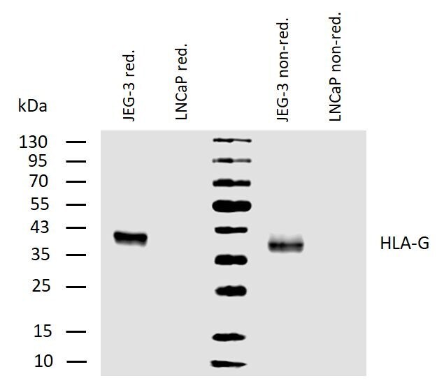
- Western blotting analysis of human HLA-G using mouse monoclonal antibody MEM-G/1 on lysates of JEG-3 cell line and LNCaP cell line (negative control) under reducing and non-reducing conditions. Nitrocellulose membrane was probed with 2µg/mL of mouse Monoclonal antibody anti-HLA-G (Product # MA1-19219) followed by IRDye800-conjugated anti-mouse secondary antibody. HLA-G was detected at approximately 40kDa.
- Submitted by
- Invitrogen Antibodies (provider)
- Main image
- Experimental details
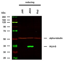
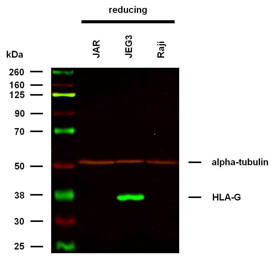
- Western Blot analysis of HLA-G using HLA-G Monoclonal Antibody (MEM-G/1) (Product # MA1-19219) under reducing conditions. Western blotting analysis was performed on whole cell extracts (RIPA lysis buffer) of JAR, JEG3, and Raji cell lines, mixed and heated (100°C, 5 min) with reducing SDS-loading buffer. Samples were resolved using 10% Tris-glycine SDS gel electrophoresis. Nitrocellulose membrane blot was probed with HLA-G Monoclonal Antibody at a dilution of 2 µg/mL, followed by IRDye 800CW Goat-anti-Mouse IgG (green). Mouse IgM anti-alpha tubulin monoclonal antibody followed by Goat-anti-Mouse IgM at a dilution of 1 µg/mL, was used as the loading control (red). Multiplex fluorescent Western blot detection was performed. HLA-G molecules were detected at ~38 kDa in JEG3 cell line.
Supportive validation
- Submitted by
- Invitrogen Antibodies (provider)
- Main image
- Experimental details
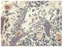
- Immunohistochemistry staining with anti-human HLA-G (MEM-G/1) Monoclonal antibody (Product # MA1-19219) - pulmonary diseases (paraffin-embedded sections). The antibody MEM-G/1 stains infiltrating macrophages in pulmonary diseases. In the top left corner see the detail of macrophage.
- Submitted by
- Invitrogen Antibodies (provider)
- Main image
- Experimental details
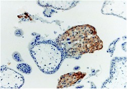
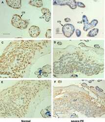
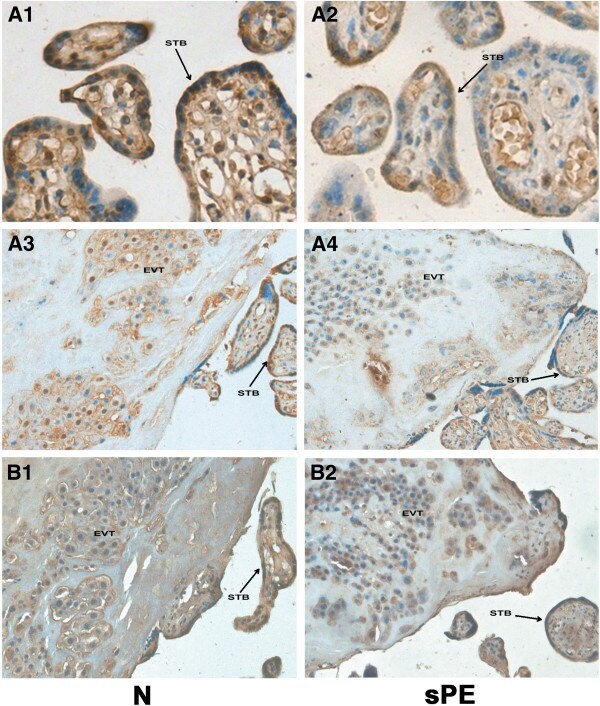
- Immunohistochemistry staining with anti-human HLA-G (MEM-G/1) Monoclonal antibody (Product # MA1-19219) of first trimester placenta (paraffin-embedded sections).

Supportive validation
- Submitted by
- Invitrogen Antibodies (provider)
- Main image

- Experimental details
- NULL
- Submitted by
- Invitrogen Antibodies (provider)
- Main image
- Experimental details
- NULL
- Submitted by
- Invitrogen Antibodies (provider)
- Main image

- Experimental details
- NULL
- Submitted by
- Invitrogen Antibodies (provider)
- Main image
- Experimental details
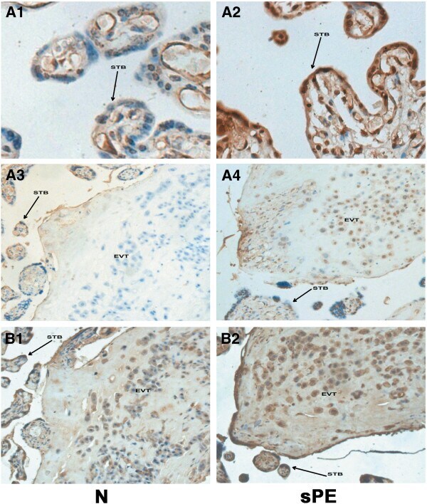
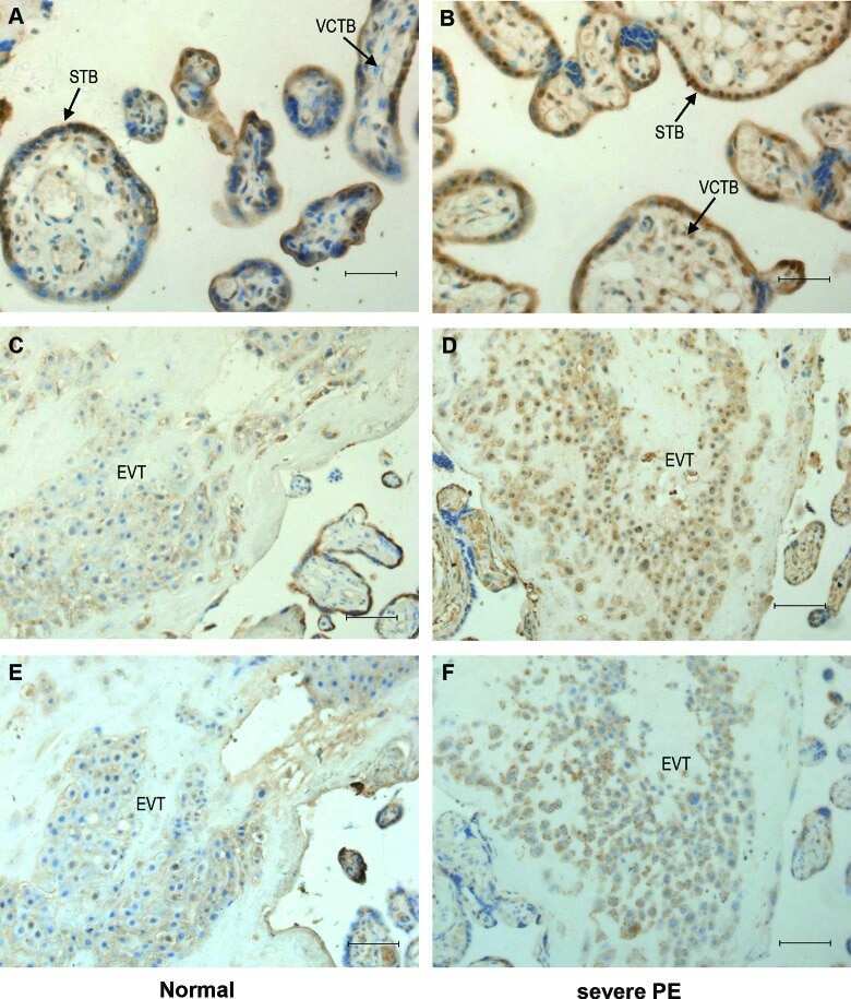
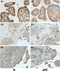
- Figure 3 Immunostaining of beta-catenin in the placental tissue sections from normal and severe preeclampsia groups. Figure 3 A1 and A3 show the immunostaining of the beta-catenin in normal control (N) group; A2 and A4 show the immunostaining of the beta-catenin in severe preeclampsia (sPE) group. The staining intensity of beta-catenin in the placental tissue of the severe PE group (A2 and A4) was weaker than the control group (A1 and A3). B1 and B2: serial sections of A3 and A4 stained with HLA-G. STB = syncytiotrophoblast, EVT = extravillous trophoblast. Original magnification: 400x for A1 and A2, 200x for A3, A4, B1, B2.
- Submitted by
- Invitrogen Antibodies (provider)
- Main image
- Experimental details
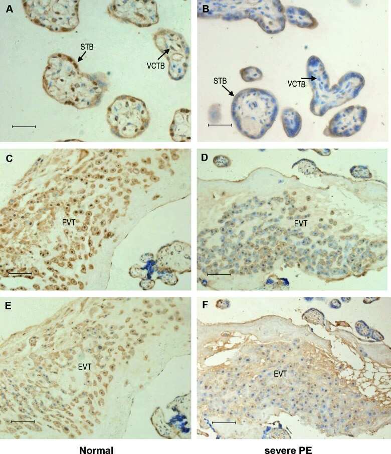
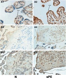
- Figure 4 Immunostaining of DKK1 in the placental tissue sections from normal and severe preeclampsia groups. Figure 4 A1 and A3 show the immunostaining of the DKK1 in normal control (N) group; A2 and A4 show the immunostaining of the DKK1 in severe preeclampsia (sPE) group. The staining intensity of DKK1 in the placental tissue of the severe PE group (A2 and A4) was greater than the control group (A1 and A3). B1 and B2: serial sections of A3 and A4 stained with HLA-G. STB = syncytiotrophoblast, EVT = extravillous trophoblast. Original magnification: 400x for A1 and A2, 200x for A3, A4, B1, B2.