Explore
 Validate
 Learn
Learn Immunocytochemistry
Antibody data
- Antibody Data
- Antigen structure
- References [1]
- Comments [0]
- Validations
- Immunocytochemistry [1]
- Immunohistochemistry [6]
- Other assay [1]
Submit
Validation data
Reference
Comment
Report error
- Product number
- PA5-53889 - Provider product page

- Provider
- Invitrogen Antibodies
- Product name
- PHLDA1 Polyclonal Antibody
- Antibody type
- Polyclonal
- Antigen
- Recombinant protein fragment
- Description
- Immunogen sequence: RMLESSGCKA LKEGVLEKRS DGLLQLWKKK CCILTEEGLL LIPPKQLQHQ QQQQQQQQQQ QQQPGQGPAE PSQPSGPAVA SLEPPVKLKE LHFSNMKTVD CVE Highest antigen sequence identity to the following orthologs: Mouse - 89%, Rat - 91%.
- Reactivity
- Human
- Host
- Rabbit
- Isotype
- IgG
- Vial size
- 100 μL
- Concentration
- 0.1 mg/mL
- Storage
- Store at 4°C short term. For long term storage, store at -20°C, avoiding freeze/thaw cycles.
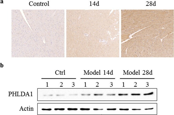
Submitted references Transcriptome Profiling Reveals PHLDA1 as a Novel Molecular Marker for Ischemic Cardiomyopathy.
Wang J, Wang F, Zhu J, Song M, An J, Li W
Journal of molecular neuroscience : MN 2018 May;65(1):102-109
Journal of molecular neuroscience : MN 2018 May;65(1):102-109
No comments: Submit comment
Supportive validation
- Submitted by
- Invitrogen Antibodies (provider)
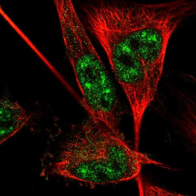
- Main image
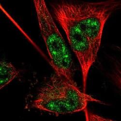
- Experimental details
- Immunofluorescent staining of PHLDA1 in human cell line U-251 MG shows positivity in nucleus but excluded from the nucleoli. Samples were probed using a PHLDA1 Polyclonal Antibody (Product # PA5-53889).
Supportive validation
- Submitted by
- Invitrogen Antibodies (provider)
- Main image
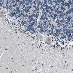
- Experimental details
- Immunohistochemical analysis of PHLDA1 in human cerebellum using PHLDA1 Polyclonal Antibody (Product # PA5-53889) shows weak cytoplasmic positivity in Purkinje cells as expected.

- Submitted by
- Invitrogen Antibodies (provider)
- Main image
- Experimental details
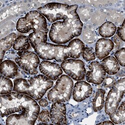
- Immunohistochemical analysis of PHLDA1 in human kidney using PHLDA1 Polyclonal Antibody (Product # PA5-53889) shows strong cytoplasmic positivity in cells in proximal tubules.

- Submitted by
- Invitrogen Antibodies (provider)
- Main image
- Experimental details
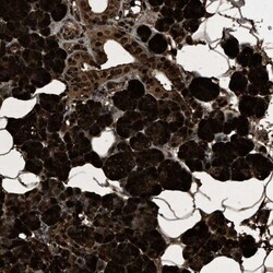
- Immunohistochemical analysis of PHLDA1 in human salivary gland using PHLDA1 Polyclonal Antibody (Product # PA5-53889) shows very strong cytoplasmic positivity in glandular cells.

- Submitted by
- Invitrogen Antibodies (provider)
- Main image

- Experimental details
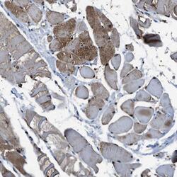
- Immunohistochemical analysis of PHLDA1 in human skeletal muscle using PHLDA1 Polyclonal Antibody (Product # PA5-53889) shows moderate cytoplasmic positivity in myocytes.

- Submitted by
- Invitrogen Antibodies (provider)
- Main image
- Experimental details
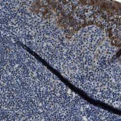
- Immunohistochemical analysis of PHLDA1 in human tonsil using PHLDA1 Polyclonal Antibody (Product # PA5-53889) shows moderate cytoplasmic positivity in squamous epithelial cells.

- Submitted by
- Invitrogen Antibodies (provider)
- Main image
- Experimental details
- Immunohistochemical analysis of PHLDA1 in human skeletal muscle using PHLDA1 Polyclonal Antibody (Product # PA5-53889) shows moderate cytoplasmic positivity in myocytes.

Supportive validation
- Submitted by
- Invitrogen Antibodies (provider)
- Main image

- Experimental details
- Fig. 2 PHLDA1 protein expression level in ICM rat model. Left ventricular tissues from control rats ( left ) or ICM rats after 14 days ( middle ) and 28 days ( right ) of coronary artery ligation were tested by immunohistochemical staining ( a ) and western blot ( b ). The result indicated increasing protein level of PHLDA1. Each group contains three rats