 Explore
 Validate
 Learn
Learn Western blot
 Immunoprecipitation
Antibody data
- Antibody Data
- Antigen structure
- References [5]
- Comments [0]
- Validations
- Western blot [2]
- Immunohistochemistry [1]
Submit
Validation data
Reference
Comment
Report error
- Product number
- NB100-68262 - Provider product page

- Provider
- Novus Biologicals
- Proper citation
- Novus Cat#NB100-68262, RRID:AB_1109819
- Product name
- Rabbit Polyclonal PHF6 Antibody
- Antibody type
- Polyclonal
- Description
- Immunogen affinity purified.
- Reactivity
- Human, Mouse
- Host
- Rabbit
- Isotype
- IgG
- Vial size
- 0.1 ml
- Concentration
- 0.2 mg/ml
- Storage
- Store at 4C. Do not freeze.
Submitted references Chromatin-Binding Protein PHF6 Regulates Activity-Dependent Transcriptional Networks to Promote Hunger Response.
PHF6 promotes non-homologous end joining and G2 checkpoint recovery.
The depletion of PHF6 decreases the drug sensitivity of T-cell acute lymphoblastic leukemia to prednisolone.
Characterization of a Mouse Model of Börjeson-Forssman-Lehmann Syndrome.
A genome-scale in vivo loss-of-function screen identifies Phf6 as a lineage-specific regulator of leukemia cell growth.
Gan L, Sun J, Yang S, Zhang X, Chen W, Sun Y, Wu X, Cheng C, Yuan J, Li A, Corbett MA, Dixon MP, Thomas T, Voss AK, Gécz J, Wang GZ, Bonni A, Li Q, Huang J
Cell reports 2020 Mar 17;30(11):3717-3728.e6
Cell reports 2020 Mar 17;30(11):3717-3728.e6
PHF6 promotes non-homologous end joining and G2 checkpoint recovery.
Warmerdam DO, Alonso-de Vega I, Wiegant WW, van den Broek B, Rother MB, Wolthuis RM, Freire R, van Attikum H, Medema RH, Smits VA
EMBO reports 2020 Jan 7;21(1):e48460
EMBO reports 2020 Jan 7;21(1):e48460
The depletion of PHF6 decreases the drug sensitivity of T-cell acute lymphoblastic leukemia to prednisolone.
Xiang J, Wang G, Xia T, Chen Z
Biomedicine & pharmacotherapy = Biomedecine & pharmacotherapie 2019 Jan;109:2210-2217
Biomedicine & pharmacotherapy = Biomedecine & pharmacotherapie 2019 Jan;109:2210-2217
Characterization of a Mouse Model of Börjeson-Forssman-Lehmann Syndrome.
Cheng C, Deng PY, Ikeuchi Y, Yuede C, Li D, Rensing N, Huang J, Baldridge D, Maloney SE, Dougherty JD, Constantino J, Jahani-Asl A, Wong M, Wozniak DF, Wang T, Klyachko VA, Bonni A
Cell reports 2018 Nov 6;25(6):1404-1414.e6
Cell reports 2018 Nov 6;25(6):1404-1414.e6
A genome-scale in vivo loss-of-function screen identifies Phf6 as a lineage-specific regulator of leukemia cell growth.
Meacham CE, Lawton LN, Soto-Feliciano YM, Pritchard JR, Joughin BA, Ehrenberger T, Fenouille N, Zuber J, Williams RT, Young RA, Hemann MT
Genes & development 2015 Mar 1;29(5):483-8
Genes & development 2015 Mar 1;29(5):483-8
No comments: Submit comment
Supportive validation
- Submitted by
- Novus Biologicals (provider)
- Main image

- Experimental details
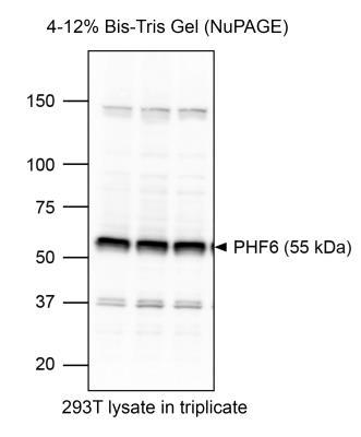
- Western Blot: PHF6 Antibody [NB100-68262] - Image from a customer review testing on 293T cells.
- Submitted by
- Novus Biologicals (provider)
- Main image

- Experimental details
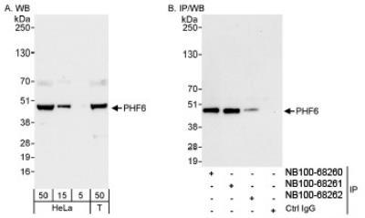
- Western Blot: PHF6 Antibody [NB100-68262] - Detection of Human PHF6 on HeLa whole cell lysate.PHF6 was also immunoprecipitated by rabbit anti-PHF6 antibodies NB100-68260 and NB100-68261.
Supportive validation
- Submitted by
- Novus Biologicals (provider)
- Main image
- Experimental details
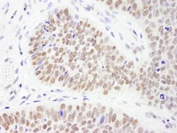
- Immunohistochemistry: PHF6 Antibody [NB100-68262] - Sample: FFPE section of human skin carcinoma. Antibody: Affinity purified rabbit anti- PHF6 used at a dilution of 1:200 (1ug/ml). Detection: DAB
