Antibody data
- Antibody Data
- Antigen structure
- References [78]
- Comments [0]
- Validations
- Immunocytochemistry [2]
- Other assay [38]
Submit
Validation data
Reference
Comment
Report error
- Product number
- A-6404 - Provider product page

- Provider
- Invitrogen Antibodies
- Product name
- MTCO2 Monoclonal Antibody (12C4F12)
- Antibody type
- Monoclonal
- Antigen
- Other
- Description
- To facilitate the study of COX structure and mitochondrial biogenesis, Invitrogen offers several subunit-specific mouse anti-OxPhos Complex IV monoclonal antibodies. The binding specificity of our anti-OxPhos Complex IV monoclonal antibody preparations allows researchers to investigate the regulation, assembly, and orientation of COX subunits in a variety of organisms. The antibodies have also proven valuable for analyzing human mitochondrial myopathies and related disorders as well as mitochondrial DNA depletion due to drug toxicity. Alexa Fluor TM 488 and Alexa Fluor TM 594 conjugates of anti- OxPhos Complex IV subunit I are also available for direct staining of these COX proteins. Cell lines can be screened for subunit expression levels for each of the OxPhos complexes by simple western blotting. These results can be combined with native gel electrophoresis or sucrose gradient centrifugation to gather additional information regarding the assembly state of the OxPhos complexes. Many of our antibodies against OxPhos subunits may also be used for immunocytochemical analysis. Image analysis of an antibody's staining pattern can reveal the relative expression and localization of a subunit. This approach has been particularly useful for studying OxPhos subunit expression in diseased muscle fibers and for screening Complex IV-deficient patients. The antibody was produced in vitro using hybridomas grown in serum-free medium, and then purified by biochemical fractionation. Near homogeneity was judged by SDS-PAGE.
- Reactivity
- Human
- Host
- Mouse
- Isotype
- IgG
- Antibody clone number
- 12C4F12
- Vial size
- 100 μg
- Concentration
- 1 mg/mL
- Storage
- 4°C, do not freeze
Submitted references The mitochondrial carrier SFXN1 is critical for complex III integrity and cellular metabolism.
SGK1 signaling promotes glucose metabolism and survival in extracellular matrix detached cells.
MTFMT deficiency correlates with reduced mitochondrial integrity and enhanced host susceptibility to intracellular infection.
Arsenic Stimulates Myoblast Mitochondrial Epidermal Growth Factor Receptor to Impair Myogenesis.
Mitochondrial Oxidative Phosphorylation Complex Regulates NLRP3 Inflammasome Activation and Predicts Patient Survival in Nasopharyngeal Carcinoma.
Differential Expression of ADP/ATP Carriers as a Biomarker of Metabolic Remodeling and Survival in Kidney Cancers.
Dissecting the Roles of Mitochondrial Complex I Intermediate Assembly Complex Factors in the Biogenesis of Complex I.
Lactic acidosis caused by repressed lactate dehydrogenase subunit B expression down-regulates mitochondrial oxidative phosphorylation via the pyruvate dehydrogenase (PDH)-PDH kinase axis.
Mitochondrial Complex I Activity Is Required for Maximal Autophagy.
Differential Alterations of the Mitochondrial Morphology and Respiratory Chain Complexes during Postnatal Development of the Mouse Lung.
Mitochondrial Maturation in Human Pluripotent Stem Cell Derived Cardiomyocytes.
Targeting mitochondrial oxidative phosphorylation eradicates therapy-resistant chronic myeloid leukemia stem cells.
Antiphospholipid antibodies increase the levels of mitochondrial DNA in placental extracellular vesicles: Alarmin-g for preeclampsia.
The human RNA-binding protein RBFA promotes the maturation of the mitochondrial ribosome.
Human adenine nucleotide translocases physically and functionally interact with respirasomes.
HIV-1 promonocytic and lymphoid cell lines: an in vitro model of in vivo mitochondrial and apoptotic lesion.
Disruption of the human COQ5-containing protein complex is associated with diminished coenzyme Q10 levels under two different conditions of mitochondrial energy deficiency.
Accessory subunits are integral for assembly and function of human mitochondrial complex I.
Improved adipose tissue function with initiation of protease inhibitor-only ART.
Melanoma addiction to the long non-coding RNA SAMMSON.
Heme Oxygenase-1/Carbon Monoxide System and Embryonic Stem Cell Differentiation and Maturation into Cardiomyocytes.
Two different pathogenic mechanisms, dying-back axonal neuropathy and pancreatic senescence, are present in the YG8R mouse model of Friedreich's ataxia.
Defective Expression of the Mitochondrial-tRNA Modifying Enzyme GTPBP3 Triggers AMPK-Mediated Adaptive Responses Involving Complex I Assembly Factors, Uncoupling Protein 2, and the Mitochondrial Pyruvate Carrier.
Loss of PTEN Facilitates Rosiglitazone-Mediated Enhancement of Platinum(IV) Complex LA-12-Induced Apoptosis in Colon Cancer Cells.
TMEM33: a new stress-inducible endoplasmic reticulum transmembrane protein and modulator of the unfolded protein response signaling.
Identification of a mitochondrial defect gene signature reveals NUPR1 as a key regulator of liver cancer progression.
Impaired Muscle Mitochondrial Biogenesis and Myogenesis in Spinal Muscular Atrophy.
A truncating PET100 variant causing fatal infantile lactic acidosis and isolated cytochrome c oxidase deficiency.
Mitochondrial Respiratory Dysfunction Induces Claudin-1 Expression via Reactive Oxygen Species-mediated Heat Shock Factor 1 Activation, Leading to Hepatoma Cell Invasiveness.
Effect of one month duration ketogenic and non-ketogenic high fat diets on mouse brain bioenergetic infrastructure.
miR-181c regulates the mitochondrial genome, bioenergetics, and propensity for heart failure in vivo.
Insulin and IGF-1 improve mitochondrial function in a PI-3K/Akt-dependent manner and reduce mitochondrial generation of reactive oxygen species in Huntington's disease knock-in striatal cells.
The ratio of Mcl-1 and Noxa determines ABT737 resistance in squamous cell carcinoma of the skin.
Evidence for mitochondrial localization of divalent metal transporter 1 (DMT1).
Mutation of the human mitochondrial phenylalanine-tRNA synthetase causes infantile-onset epilepsy and cytochrome c oxidase deficiency.
Glycogen synthase kinase-3 (GSK3) controls deoxyglucose-induced mitochondrial biogenesis in human neuroblastoma SH-SY5Y cells.
Deoxypyrimidine monophosphate bypass therapy for thymidine kinase 2 deficiency.
Ridaifen-SB8, a novel tamoxifen derivative, induces apoptosis via reactive oxygen species-dependent signaling pathway.
Mitochondria and tumor progression in ulcerative colitis.
Identification of elongation factor G as the conserved cellular target of argyrin B.
Pilot application of iTRAQ to the retinal disease Macular Telangiectasia.
Mutations in MTFMT underlie a human disorder of formylation causing impaired mitochondrial translation.
Mitochondrial proteins, learning and memory: biochemical specialization of a memory system.
LMNA mutations induce a non-inflammatory fibrosis and a brown fat-like dystrophy of enlarged cervical adipose tissue.
Cyclin D1 inhibits mitochondrial activity in B cells.
Human ERAL1 is a mitochondrial RNA chaperone involved in the assembly of the 28S small mitochondrial ribosomal subunit.
Altered gene transcription profiles in fibroblasts harboring either TK2 or DGUOK mutations indicate compensatory mechanisms.
PUMA- and Bax-induced autophagy contributes to apoptosis.
Dysregulation of mitochondrial biogenesis in vascular endothelial and smooth muscle cells of aged rats.
A novel histone deacetylase 8 (HDAC8)-specific inhibitor PCI-34051 induces apoptosis in T-cell lymphomas.
Perioculomotor cell groups in monkey and man defined by their histochemical and functional properties: reappraisal of the Edinger-Westphal nucleus.
Human lipodystrophies linked to mutations in A-type lamins and to HIV protease inhibitor therapy are both associated with prelamin A accumulation, oxidative stress and premature cellular senescence.
Identification of mouse Prp19p as a lipid droplet-associated protein and its possible involvement in the biogenesis of lipid droplets.
Coordination of nuclear- and mitochondrial-DNA encoded proteins in cancer and normal colon tissues.
Mcl-1 interacts with truncated Bid and inhibits its induction of cytochrome c release and its role in receptor-mediated apoptosis.
Differential expression proteomics of human colon cancer.
Localization of mitochondrial DNA encoded cytochrome c oxidase subunits I and II in rat pancreatic zymogen granules and pituitary growth hormone granules.
Tissue-specific cytochrome c oxidase assembly defects due to mutations in SCO2 and SURF1.
Heat shock pretreatment inhibited the release of Smac/DIABLO from mitochondria and apoptosis induced by hydrogen peroxide in cardiomyocytes and C2C12 myogenic cells.
Oxidative stress and the mitochondrial theory of aging in human skeletal muscle.
Tim50, a component of the mitochondrial translocator, regulates mitochondrial integrity and cell death.
Lactacystin-induced apoptosis of cultured mouse cortical neurons is associated with accumulation of PTEN in the detergent-resistant membrane fraction.
Cotreatment with histone deacetylase inhibitor LAQ824 enhances Apo-2L/tumor necrosis factor-related apoptosis inducing ligand-induced death inducing signaling complex activity and apoptosis of human acute leukemia cells.
Mitochondrially localized active caspase-9 and caspase-3 result mostly from translocation from the cytosol and partly from caspase-mediated activation in the organelle. Lack of evidence for Apaf-1-mediated procaspase-9 activation in the mitochondria.
Bisindolylmaleimide IX facilitates tumor necrosis factor receptor family-mediated cell death and acts as an inhibitor of transcription.
Differential cytostatic and apoptotic effects of ecteinascidin-743 in cancer cells. Transcription-dependent cell cycle arrest and transcription-independent JNK and mitochondrial mediated apoptosis.
Early mitochondrial activation and cytochrome c up-regulation during apoptosis.
Caspase-2 induces apoptosis by releasing proapoptotic proteins from mitochondria.
Bid, a widely expressed proapoptotic protein of the Bcl-2 family, displays lipid transfer activity.
Role of mitochondria and caspases in vitamin D-mediated apoptosis of MCF-7 breast cancer cells.
The chaperone function of hsp70 is required for protection against stress-induced apoptosis.
The chaperone function of hsp70 is required for protection against stress-induced apoptosis.
Tauroursodeoxycholic acid partially prevents apoptosis induced by 3-nitropropionic acid: evidence for a mitochondrial pathway independent of the permeability transition.
TID1, a human homolog of the Drosophila tumor suppressor l(2)tid, encodes two mitochondrial modulators of apoptosis with opposing functions.
TID1, a human homolog of the Drosophila tumor suppressor l(2)tid, encodes two mitochondrial modulators of apoptosis with opposing functions.
Dual targeting property of the N-terminal signal sequence of P4501A1. Targeting of heterologous proteins to endoplasmic reticulum and mitochondria.
Mammalian cytochrome-c oxidase: characterization of enzyme and immunological detection of subunits in tissue extracts and whole cells.
Mammalian cytochrome-c oxidase: characterization of enzyme and immunological detection of subunits in tissue extracts and whole cells.
Acoba MG, Alpergin ESS, Renuse S, Fernández-Del-Río L, Lu YW, Khalimonchuk O, Clarke CF, Pandey A, Wolfgang MJ, Claypool SM
Cell reports 2021 Mar 16;34(11):108869
Cell reports 2021 Mar 16;34(11):108869
SGK1 signaling promotes glucose metabolism and survival in extracellular matrix detached cells.
Mason JA, Cockfield JA, Pape DJ, Meissner H, Sokolowski MT, White TC, Valentín López JC, Liu J, Liu X, Martínez-Reyes I, Chandel NS, Locasale JW, Schafer ZT
Cell reports 2021 Mar 16;34(11):108821
Cell reports 2021 Mar 16;34(11):108821
MTFMT deficiency correlates with reduced mitochondrial integrity and enhanced host susceptibility to intracellular infection.
Seo JH, Hwang CS, Yoo JY
Scientific reports 2020 Jul 7;10(1):11183
Scientific reports 2020 Jul 7;10(1):11183
Arsenic Stimulates Myoblast Mitochondrial Epidermal Growth Factor Receptor to Impair Myogenesis.
Cheikhi A, Anguiano T, Lasak J, Qian B, Sahu A, Mimiya H, Cohen CC, Wipf P, Ambrosio F, Barchowsky A
Toxicological sciences : an official journal of the Society of Toxicology 2020 Jul 1;176(1):162-174
Toxicological sciences : an official journal of the Society of Toxicology 2020 Jul 1;176(1):162-174
Mitochondrial Oxidative Phosphorylation Complex Regulates NLRP3 Inflammasome Activation and Predicts Patient Survival in Nasopharyngeal Carcinoma.
Chung IC, Chen LC, Tsang NM, Chuang WY, Liao TC, Yuan SN, OuYang CN, Ojcius DM, Wu CC, Chang YS
Molecular & cellular proteomics : MCP 2020 Jan;19(1):142-154
Molecular & cellular proteomics : MCP 2020 Jan;19(1):142-154
Differential Expression of ADP/ATP Carriers as a Biomarker of Metabolic Remodeling and Survival in Kidney Cancers.
Trisolini L, Laera L, Favia M, Muscella A, Castegna A, Pesce V, Guerra L, De Grassi A, Volpicella M, Pierri CL
Biomolecules 2020 Dec 30;11(1)
Biomolecules 2020 Dec 30;11(1)
Dissecting the Roles of Mitochondrial Complex I Intermediate Assembly Complex Factors in the Biogenesis of Complex I.
Formosa LE, Muellner-Wong L, Reljic B, Sharpe AJ, Jackson TD, Beilharz TH, Stojanovski D, Lazarou M, Stroud DA, Ryan MT
Cell reports 2020 Apr 21;31(3):107541
Cell reports 2020 Apr 21;31(3):107541
Lactic acidosis caused by repressed lactate dehydrogenase subunit B expression down-regulates mitochondrial oxidative phosphorylation via the pyruvate dehydrogenase (PDH)-PDH kinase axis.
Hong SM, Lee YK, Park I, Kwon SM, Min S, Yoon G
The Journal of biological chemistry 2019 May 10;294(19):7810-7820
The Journal of biological chemistry 2019 May 10;294(19):7810-7820
Mitochondrial Complex I Activity Is Required for Maximal Autophagy.
Thomas HE, Zhang Y, Stefely JA, Veiga SR, Thomas G, Kozma SC, Mercer CA
Cell reports 2018 Aug 28;24(9):2404-2417.e8
Cell reports 2018 Aug 28;24(9):2404-2417.e8
Differential Alterations of the Mitochondrial Morphology and Respiratory Chain Complexes during Postnatal Development of the Mouse Lung.
El-Merhie N, Baumgart-Vogt E, Pilatz A, Pfreimer S, Pfeiffer B, Pak O, Kosanovic D, Seimetz M, Schermuly RT, Weissmann N, Karnati S
Oxidative medicine and cellular longevity 2017;2017:9169146
Oxidative medicine and cellular longevity 2017;2017:9169146
Mitochondrial Maturation in Human Pluripotent Stem Cell Derived Cardiomyocytes.
Dai DF, Danoviz ME, Wiczer B, Laflamme MA, Tian R
Stem cells international 2017;2017:5153625
Stem cells international 2017;2017:5153625
Targeting mitochondrial oxidative phosphorylation eradicates therapy-resistant chronic myeloid leukemia stem cells.
Kuntz EM, Baquero P, Michie AM, Dunn K, Tardito S, Holyoake TL, Helgason GV, Gottlieb E
Nature medicine 2017 Oct;23(10):1234-1240
Nature medicine 2017 Oct;23(10):1234-1240
Antiphospholipid antibodies increase the levels of mitochondrial DNA in placental extracellular vesicles: Alarmin-g for preeclampsia.
Tong M, Johansson C, Xiao F, Stone PR, James JL, Chen Q, Cree LM, Chamley LW
Scientific reports 2017 Nov 29;7(1):16556
Scientific reports 2017 Nov 29;7(1):16556
The human RNA-binding protein RBFA promotes the maturation of the mitochondrial ribosome.
Rozanska A, Richter-Dennerlein R, Rorbach J, Gao F, Lewis RJ, Chrzanowska-Lightowlers ZM, Lightowlers RN
The Biochemical journal 2017 Jun 13;474(13):2145-2158
The Biochemical journal 2017 Jun 13;474(13):2145-2158
Human adenine nucleotide translocases physically and functionally interact with respirasomes.
Lu YW, Acoba MG, Selvaraju K, Huang TC, Nirujogi RS, Sathe G, Pandey A, Claypool SM
Molecular biology of the cell 2017 Jun 1;28(11):1489-1506
Molecular biology of the cell 2017 Jun 1;28(11):1489-1506
HIV-1 promonocytic and lymphoid cell lines: an in vitro model of in vivo mitochondrial and apoptotic lesion.
Morén C, González-Casacuberta I, Álvarez-Fernández C, Bañó M, Catalán-Garcia M, Guitart-Mampel M, Juárez-Flores DL, Tobías E, Milisenda J, Cardellach F, Gatell JM, Sánchez-Palomino S, Garrabou G
Journal of cellular and molecular medicine 2017 Feb;21(2):402-409
Journal of cellular and molecular medicine 2017 Feb;21(2):402-409
Disruption of the human COQ5-containing protein complex is associated with diminished coenzyme Q10 levels under two different conditions of mitochondrial energy deficiency.
Yen HC, Liu YC, Kan CC, Wei HJ, Lee SH, Wei YH, Feng YH, Chen CW, Huang CC
Biochimica et biophysica acta 2016 Sep;1860(9):1864-76
Biochimica et biophysica acta 2016 Sep;1860(9):1864-76
Accessory subunits are integral for assembly and function of human mitochondrial complex I.
Stroud DA, Surgenor EE, Formosa LE, Reljic B, Frazier AE, Dibley MG, Osellame LD, Stait T, Beilharz TH, Thorburn DR, Salim A, Ryan MT
Nature 2016 Oct 6;538(7623):123-126
Nature 2016 Oct 6;538(7623):123-126
Improved adipose tissue function with initiation of protease inhibitor-only ART.
Maughan RT, Feeney ER, Capel E, Capeau J, Domingo P, Giralt M, Lange JM, Phanuphak P, Cooper DA, Reiss P, Mallon PW, HIVNAT-019 Study Group
The Journal of antimicrobial chemotherapy 2016 Nov;71(11):3212-3221
The Journal of antimicrobial chemotherapy 2016 Nov;71(11):3212-3221
Melanoma addiction to the long non-coding RNA SAMMSON.
Leucci E, Vendramin R, Spinazzi M, Laurette P, Fiers M, Wouters J, Radaelli E, Eyckerman S, Leonelli C, Vanderheyden K, Rogiers A, Hermans E, Baatsen P, Aerts S, Amant F, Van Aelst S, van den Oord J, de Strooper B, Davidson I, Lafontaine DL, Gevaert K, Vandesompele J, Mestdagh P, Marine JC
Nature 2016 Mar 24;531(7595):518-22
Nature 2016 Mar 24;531(7595):518-22
Heme Oxygenase-1/Carbon Monoxide System and Embryonic Stem Cell Differentiation and Maturation into Cardiomyocytes.
Suliman HB, Zobi F, Piantadosi CA
Antioxidants & redox signaling 2016 Mar 1;24(7):345-60
Antioxidants & redox signaling 2016 Mar 1;24(7):345-60
Two different pathogenic mechanisms, dying-back axonal neuropathy and pancreatic senescence, are present in the YG8R mouse model of Friedreich's ataxia.
Mollá B, Riveiro F, Bolinches-Amorós A, Muñoz-Lasso DC, Palau F, González-Cabo P
Disease models & mechanisms 2016 Jun 1;9(6):647-57
Disease models & mechanisms 2016 Jun 1;9(6):647-57
Defective Expression of the Mitochondrial-tRNA Modifying Enzyme GTPBP3 Triggers AMPK-Mediated Adaptive Responses Involving Complex I Assembly Factors, Uncoupling Protein 2, and the Mitochondrial Pyruvate Carrier.
Martínez-Zamora A, Meseguer S, Esteve JM, Villarroya M, Aguado C, Enríquez JA, Knecht E, Armengod ME
PloS one 2015;10(12):e0144273
PloS one 2015;10(12):e0144273
Loss of PTEN Facilitates Rosiglitazone-Mediated Enhancement of Platinum(IV) Complex LA-12-Induced Apoptosis in Colon Cancer Cells.
Lauková J, Kozubík A, Hofmanová J, Nekvindová J, Sova P, Moyer MP, Ehrmann J, Hyršlová Vaculová A
PloS one 2015;10(10):e0141020
PloS one 2015;10(10):e0141020
TMEM33: a new stress-inducible endoplasmic reticulum transmembrane protein and modulator of the unfolded protein response signaling.
Sakabe I, Hu R, Jin L, Clarke R, Kasid UN
Breast cancer research and treatment 2015 Sep;153(2):285-97
Breast cancer research and treatment 2015 Sep;153(2):285-97
Identification of a mitochondrial defect gene signature reveals NUPR1 as a key regulator of liver cancer progression.
Lee YK, Jee BA, Kwon SM, Yoon YS, Xu WG, Wang HJ, Wang XW, Thorgeirsson SS, Lee JS, Woo HG, Yoon G
Hepatology (Baltimore, Md.) 2015 Oct;62(4):1174-89
Hepatology (Baltimore, Md.) 2015 Oct;62(4):1174-89
Impaired Muscle Mitochondrial Biogenesis and Myogenesis in Spinal Muscular Atrophy.
Ripolone M, Ronchi D, Violano R, Vallejo D, Fagiolari G, Barca E, Lucchini V, Colombo I, Villa L, Berardinelli A, Balottin U, Morandi L, Mora M, Bordoni A, Fortunato F, Corti S, Parisi D, Toscano A, Sciacco M, DiMauro S, Comi GP, Moggio M
JAMA neurology 2015 Jun;72(6):666-75
JAMA neurology 2015 Jun;72(6):666-75
A truncating PET100 variant causing fatal infantile lactic acidosis and isolated cytochrome c oxidase deficiency.
Oláhová M, Haack TB, Alston CL, Houghton JA, He L, Morris AA, Brown GK, McFarland R, Chrzanowska-Lightowlers ZM, Lightowlers RN, Prokisch H, Taylor RW
European journal of human genetics : EJHG 2015 Jul;23(7):935-9
European journal of human genetics : EJHG 2015 Jul;23(7):935-9
Mitochondrial Respiratory Dysfunction Induces Claudin-1 Expression via Reactive Oxygen Species-mediated Heat Shock Factor 1 Activation, Leading to Hepatoma Cell Invasiveness.
Lee JH, Lee YK, Lim JJ, Byun HO, Park I, Kim GH, Xu WG, Wang HJ, Yoon G
The Journal of biological chemistry 2015 Aug 28;290(35):21421-31
The Journal of biological chemistry 2015 Aug 28;290(35):21421-31
Effect of one month duration ketogenic and non-ketogenic high fat diets on mouse brain bioenergetic infrastructure.
Selfridge JE, Wilkins HM, E L, Carl SM, Koppel S, Funk E, Fields T, Lu J, Tang EP, Slawson C, Wang W, Zhu H, Swerdlow RH
Journal of bioenergetics and biomembranes 2015 Apr;47(1-2):1-11
Journal of bioenergetics and biomembranes 2015 Apr;47(1-2):1-11
miR-181c regulates the mitochondrial genome, bioenergetics, and propensity for heart failure in vivo.
Das S, Bedja D, Campbell N, Dunkerly B, Chenna V, Maitra A, Steenbergen C
PloS one 2014;9(5):e96820
PloS one 2014;9(5):e96820
Insulin and IGF-1 improve mitochondrial function in a PI-3K/Akt-dependent manner and reduce mitochondrial generation of reactive oxygen species in Huntington's disease knock-in striatal cells.
Ribeiro M, Rosenstock TR, Oliveira AM, Oliveira CR, Rego AC
Free radical biology & medicine 2014 Sep;74:129-44
Free radical biology & medicine 2014 Sep;74:129-44
The ratio of Mcl-1 and Noxa determines ABT737 resistance in squamous cell carcinoma of the skin.
Geserick P, Wang J, Feoktistova M, Leverkus M
Cell death & disease 2014 Sep 11;5(9):e1412
Cell death & disease 2014 Sep 11;5(9):e1412
Evidence for mitochondrial localization of divalent metal transporter 1 (DMT1).
Wolff NA, Ghio AJ, Garrick LM, Garrick MD, Zhao L, Fenton RA, Thévenod F
FASEB journal : official publication of the Federation of American Societies for Experimental Biology 2014 May;28(5):2134-45
FASEB journal : official publication of the Federation of American Societies for Experimental Biology 2014 May;28(5):2134-45
Mutation of the human mitochondrial phenylalanine-tRNA synthetase causes infantile-onset epilepsy and cytochrome c oxidase deficiency.
Almalki A, Alston CL, Parker A, Simonic I, Mehta SG, He L, Reza M, Oliveira JM, Lightowlers RN, McFarland R, Taylor RW, Chrzanowska-Lightowlers ZM
Biochimica et biophysica acta 2014 Jan;1842(1):56-64
Biochimica et biophysica acta 2014 Jan;1842(1):56-64
Glycogen synthase kinase-3 (GSK3) controls deoxyglucose-induced mitochondrial biogenesis in human neuroblastoma SH-SY5Y cells.
Ngamsiri P, Watcharasit P, Satayavivad J
Mitochondrion 2014 Jan;14(1):54-63
Mitochondrion 2014 Jan;14(1):54-63
Deoxypyrimidine monophosphate bypass therapy for thymidine kinase 2 deficiency.
Garone C, Garcia-Diaz B, Emmanuele V, Lopez LC, Tadesse S, Akman HO, Tanji K, Quinzii CM, Hirano M
EMBO molecular medicine 2014 Aug;6(8):1016-27
EMBO molecular medicine 2014 Aug;6(8):1016-27
Ridaifen-SB8, a novel tamoxifen derivative, induces apoptosis via reactive oxygen species-dependent signaling pathway.
Guo WZ, Shiina I, Wang Y, Umeda E, Watanabe C, Uetake S, Ohashi Y, Yamori T, Dan S
Biochemical pharmacology 2013 Nov 1;86(9):1272-84
Biochemical pharmacology 2013 Nov 1;86(9):1272-84
Mitochondria and tumor progression in ulcerative colitis.
Ussakli CH, Ebaee A, Binkley J, Brentnall TA, Emond MJ, Rabinovitch PS, Risques RA
Journal of the National Cancer Institute 2013 Aug 21;105(16):1239-48
Journal of the National Cancer Institute 2013 Aug 21;105(16):1239-48
Identification of elongation factor G as the conserved cellular target of argyrin B.
Nyfeler B, Hoepfner D, Palestrant D, Kirby CA, Whitehead L, Yu R, Deng G, Caughlan RE, Woods AL, Jones AK, Barnes SW, Walker JR, Gaulis S, Hauy E, Brachmann SM, Krastel P, Studer C, Riedl R, Estoppey D, Aust T, Movva NR, Wang Z, Salcius M, Michaud GA, McAllister G, Murphy LO, Tallarico JA, Wilson CJ, Dean CR
PloS one 2012;7(9):e42657
PloS one 2012;7(9):e42657
Pilot application of iTRAQ to the retinal disease Macular Telangiectasia.
Len AC, Powner MB, Zhu L, Hageman GS, Song X, Fruttiger M, Gillies MC
Journal of proteome research 2012 Feb 3;11(2):537-53
Journal of proteome research 2012 Feb 3;11(2):537-53
Mutations in MTFMT underlie a human disorder of formylation causing impaired mitochondrial translation.
Tucker EJ, Hershman SG, Köhrer C, Belcher-Timme CA, Patel J, Goldberger OA, Christodoulou J, Silberstein JM, McKenzie M, Ryan MT, Compton AG, Jaffe JD, Carr SA, Calvo SE, RajBhandary UL, Thorburn DR, Mootha VK
Cell metabolism 2011 Sep 7;14(3):428-34
Cell metabolism 2011 Sep 7;14(3):428-34
Mitochondrial proteins, learning and memory: biochemical specialization of a memory system.
Solomonia RO, Kunelauri N, Mikautadze E, Apkhazava D, McCabe BJ, Horn G
Neuroscience 2011 Oct 27;194:112-23
Neuroscience 2011 Oct 27;194:112-23
LMNA mutations induce a non-inflammatory fibrosis and a brown fat-like dystrophy of enlarged cervical adipose tissue.
Béréziat V, Cervera P, Le Dour C, Verpont MC, Dumont S, Vantyghem MC, Capeau J, Vigouroux C, Lipodystrophy Study Group
The American journal of pathology 2011 Nov;179(5):2443-53
The American journal of pathology 2011 Nov;179(5):2443-53
Cyclin D1 inhibits mitochondrial activity in B cells.
Tchakarska G, Roussel M, Troussard X, Sola B
Cancer research 2011 Mar 1;71(5):1690-9
Cancer research 2011 Mar 1;71(5):1690-9
Human ERAL1 is a mitochondrial RNA chaperone involved in the assembly of the 28S small mitochondrial ribosomal subunit.
Dennerlein S, Rozanska A, Wydro M, Chrzanowska-Lightowlers ZM, Lightowlers RN
The Biochemical journal 2010 Sep 15;430(3):551-8
The Biochemical journal 2010 Sep 15;430(3):551-8
Altered gene transcription profiles in fibroblasts harboring either TK2 or DGUOK mutations indicate compensatory mechanisms.
Villarroya J, de Bolós C, Meseguer A, Hirano M, Vilà MR
Experimental cell research 2009 May 1;315(8):1429-38
Experimental cell research 2009 May 1;315(8):1429-38
PUMA- and Bax-induced autophagy contributes to apoptosis.
Yee KS, Wilkinson S, James J, Ryan KM, Vousden KH
Cell death and differentiation 2009 Aug;16(8):1135-45
Cell death and differentiation 2009 Aug;16(8):1135-45
Dysregulation of mitochondrial biogenesis in vascular endothelial and smooth muscle cells of aged rats.
Ungvari Z, Labinskyy N, Gupte S, Chander PN, Edwards JG, Csiszar A
American journal of physiology. Heart and circulatory physiology 2008 May;294(5):H2121-8
American journal of physiology. Heart and circulatory physiology 2008 May;294(5):H2121-8
A novel histone deacetylase 8 (HDAC8)-specific inhibitor PCI-34051 induces apoptosis in T-cell lymphomas.
Balasubramanian S, Ramos J, Luo W, Sirisawad M, Verner E, Buggy JJ
Leukemia 2008 May;22(5):1026-34
Leukemia 2008 May;22(5):1026-34
Perioculomotor cell groups in monkey and man defined by their histochemical and functional properties: reappraisal of the Edinger-Westphal nucleus.
Horn AK, Eberhorn A, Härtig W, Ardeleanu P, Messoudi A, Büttner-Ennever JA
The Journal of comparative neurology 2008 Mar 20;507(3):1317-35
The Journal of comparative neurology 2008 Mar 20;507(3):1317-35
Human lipodystrophies linked to mutations in A-type lamins and to HIV protease inhibitor therapy are both associated with prelamin A accumulation, oxidative stress and premature cellular senescence.
Caron M, Auclair M, Donadille B, Béréziat V, Guerci B, Laville M, Narbonne H, Bodemer C, Lascols O, Capeau J, Vigouroux C
Cell death and differentiation 2007 Oct;14(10):1759-67
Cell death and differentiation 2007 Oct;14(10):1759-67
Identification of mouse Prp19p as a lipid droplet-associated protein and its possible involvement in the biogenesis of lipid droplets.
Cho SY, Shin ES, Park PJ, Shin DW, Chang HK, Kim D, Lee HH, Lee JH, Kim SH, Song MJ, Chang IS, Lee OS, Lee TR
The Journal of biological chemistry 2007 Jan 26;282(4):2456-65
The Journal of biological chemistry 2007 Jan 26;282(4):2456-65
Coordination of nuclear- and mitochondrial-DNA encoded proteins in cancer and normal colon tissues.
Mazzanti R, Giulivi C
Biochimica et biophysica acta 2006 May-Jun;1757(5-6):618-23
Biochimica et biophysica acta 2006 May-Jun;1757(5-6):618-23
Mcl-1 interacts with truncated Bid and inhibits its induction of cytochrome c release and its role in receptor-mediated apoptosis.
Clohessy JG, Zhuang J, de Boer J, Gil-Gómez G, Brady HJ
The Journal of biological chemistry 2006 Mar 3;281(9):5750-9
The Journal of biological chemistry 2006 Mar 3;281(9):5750-9
Differential expression proteomics of human colon cancer.
Mazzanti R, Solazzo M, Fantappié O, Elfering S, Pantaleo P, Bechi P, Cianchi F, Ettl A, Giulivi C
American journal of physiology. Gastrointestinal and liver physiology 2006 Jun;290(6):G1329-38
American journal of physiology. Gastrointestinal and liver physiology 2006 Jun;290(6):G1329-38
Localization of mitochondrial DNA encoded cytochrome c oxidase subunits I and II in rat pancreatic zymogen granules and pituitary growth hormone granules.
Sadacharan SK, Singh B, Bowes T, Gupta RS
Histochemistry and cell biology 2005 Nov;124(5):409-21
Histochemistry and cell biology 2005 Nov;124(5):409-21
Tissue-specific cytochrome c oxidase assembly defects due to mutations in SCO2 and SURF1.
Stiburek L, Vesela K, Hansikova H, Pecina P, Tesarova M, Cerna L, Houstek J, Zeman J
The Biochemical journal 2005 Dec 15;392(Pt 3):625-32
The Biochemical journal 2005 Dec 15;392(Pt 3):625-32
Heat shock pretreatment inhibited the release of Smac/DIABLO from mitochondria and apoptosis induced by hydrogen peroxide in cardiomyocytes and C2C12 myogenic cells.
Jiang B, Xiao W, Shi Y, Liu M, Xiao X
Cell stress & chaperones 2005 Autumn;10(3):252-62
Cell stress & chaperones 2005 Autumn;10(3):252-62
Oxidative stress and the mitochondrial theory of aging in human skeletal muscle.
Gianni P, Jan KJ, Douglas MJ, Stuart PM, Tarnopolsky MA
Experimental gerontology 2004 Sep;39(9):1391-400
Experimental gerontology 2004 Sep;39(9):1391-400
Tim50, a component of the mitochondrial translocator, regulates mitochondrial integrity and cell death.
Guo Y, Cheong N, Zhang Z, De Rose R, Deng Y, Farber SA, Fernandes-Alnemri T, Alnemri ES
The Journal of biological chemistry 2004 Jun 4;279(23):24813-25
The Journal of biological chemistry 2004 Jun 4;279(23):24813-25
Lactacystin-induced apoptosis of cultured mouse cortical neurons is associated with accumulation of PTEN in the detergent-resistant membrane fraction.
Cheung NS, Choy MS, Halliwell B, Teo TS, Bay BH, Lee AY, Qi RZ, Koh VH, Whiteman M, Koay ES, Chiu LL, Zhu HJ, Wong KP, Beart PM, Cheng HC
Cellular and molecular life sciences : CMLS 2004 Aug;61(15):1926-34
Cellular and molecular life sciences : CMLS 2004 Aug;61(15):1926-34
Cotreatment with histone deacetylase inhibitor LAQ824 enhances Apo-2L/tumor necrosis factor-related apoptosis inducing ligand-induced death inducing signaling complex activity and apoptosis of human acute leukemia cells.
Guo F, Sigua C, Tao J, Bali P, George P, Li Y, Wittmann S, Moscinski L, Atadja P, Bhalla K
Cancer research 2004 Apr 1;64(7):2580-9
Cancer research 2004 Apr 1;64(7):2580-9
Mitochondrially localized active caspase-9 and caspase-3 result mostly from translocation from the cytosol and partly from caspase-mediated activation in the organelle. Lack of evidence for Apaf-1-mediated procaspase-9 activation in the mitochondria.
Chandra D, Tang DG
The Journal of biological chemistry 2003 May 9;278(19):17408-20
The Journal of biological chemistry 2003 May 9;278(19):17408-20
Bisindolylmaleimide IX facilitates tumor necrosis factor receptor family-mediated cell death and acts as an inhibitor of transcription.
Rokhlin OW, Glover RA, Taghiyev AF, Guseva NV, Seftor RE, Shyshynova I, Gudkov AV, Cohen MB
The Journal of biological chemistry 2002 Sep 6;277(36):33213-9
The Journal of biological chemistry 2002 Sep 6;277(36):33213-9
Differential cytostatic and apoptotic effects of ecteinascidin-743 in cancer cells. Transcription-dependent cell cycle arrest and transcription-independent JNK and mitochondrial mediated apoptosis.
Gajate C, An F, Mollinedo F
The Journal of biological chemistry 2002 Nov 1;277(44):41580-9
The Journal of biological chemistry 2002 Nov 1;277(44):41580-9
Early mitochondrial activation and cytochrome c up-regulation during apoptosis.
Chandra D, Liu JW, Tang DG
The Journal of biological chemistry 2002 Dec 27;277(52):50842-54
The Journal of biological chemistry 2002 Dec 27;277(52):50842-54
Caspase-2 induces apoptosis by releasing proapoptotic proteins from mitochondria.
Guo Y, Srinivasula SM, Druilhe A, Fernandes-Alnemri T, Alnemri ES
The Journal of biological chemistry 2002 Apr 19;277(16):13430-7
The Journal of biological chemistry 2002 Apr 19;277(16):13430-7
Bid, a widely expressed proapoptotic protein of the Bcl-2 family, displays lipid transfer activity.
Esposti MD, Erler JT, Hickman JA, Dive C
Molecular and cellular biology 2001 Nov;21(21):7268-76
Molecular and cellular biology 2001 Nov;21(21):7268-76
Role of mitochondria and caspases in vitamin D-mediated apoptosis of MCF-7 breast cancer cells.
Narvaez CJ, Welsh J
The Journal of biological chemistry 2001 Mar 23;276(12):9101-7
The Journal of biological chemistry 2001 Mar 23;276(12):9101-7
The chaperone function of hsp70 is required for protection against stress-induced apoptosis.
Mosser DD, Caron AW, Bourget L, Meriin AB, Sherman MY, Morimoto RI, Massie B
Molecular and cellular biology 2000 Oct;20(19):7146-59
Molecular and cellular biology 2000 Oct;20(19):7146-59
The chaperone function of hsp70 is required for protection against stress-induced apoptosis.
Mosser DD, Caron AW, Bourget L, Meriin AB, Sherman MY, Morimoto RI, Massie B
Molecular and cellular biology 2000 Oct;20(19):7146-59
Molecular and cellular biology 2000 Oct;20(19):7146-59
Tauroursodeoxycholic acid partially prevents apoptosis induced by 3-nitropropionic acid: evidence for a mitochondrial pathway independent of the permeability transition.
Rodrigues CM, Stieers CL, Keene CD, Ma X, Kren BT, Low WC, Steer CJ
Journal of neurochemistry 2000 Dec;75(6):2368-79
Journal of neurochemistry 2000 Dec;75(6):2368-79
TID1, a human homolog of the Drosophila tumor suppressor l(2)tid, encodes two mitochondrial modulators of apoptosis with opposing functions.
Syken J, De-Medina T, Münger K
Proceedings of the National Academy of Sciences of the United States of America 1999 Jul 20;96(15):8499-504
Proceedings of the National Academy of Sciences of the United States of America 1999 Jul 20;96(15):8499-504
Dual targeting property of the N-terminal signal sequence of P4501A1. Targeting of heterologous proteins to endoplasmic reticulum and mitochondria.
Bhagwat SV, Biswas G, Anandatheerthavarada HK, Addya S, Pandak W, Avadhani NG
The Journal of biological chemistry 1999 Aug 20;274(34):24014-22
The Journal of biological chemistry 1999 Aug 20;274(34):24014-22
Mammalian cytochrome-c oxidase: characterization of enzyme and immunological detection of subunits in tissue extracts and whole cells.
Capaldi RA, Marusich MF, Taanman JW
Methods in enzymology 1995;260:117-32
Methods in enzymology 1995;260:117-32
Mammalian cytochrome-c oxidase: characterization of enzyme and immunological detection of subunits in tissue extracts and whole cells.
Capaldi RA, Marusich MF, Taanman JW
Methods in enzymology 1995;260:117-32
Methods in enzymology 1995;260:117-32
No comments: Submit comment
Supportive validation
- Submitted by
- Invitrogen Antibodies (provider)
- Main image
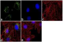
- Experimental details
- Immunofluorescence analysis of OXPHOS COMPLEX IV SU (MTCO2) was performed using 90% confluent log phase Hep G2 cells. The cells were fixed with 4% paraformaldehyde for 10 minutes, permeabilized with 0.1% Triton™ X-100 for 10 minutes, and blocked with 1% BSA for 1 hour at room temperature. The cells were labeled with MTCO2 (12C4F12) Mouse Monoclonal Antibody (Product # A-6404) at 2µg/mL in 0.1% BSA and incubated for 3 hours at room temperature and then labeled with Goat anti-Mouse IgG (H+L) Superclonal™ Secondary Antibody, Alexa Fluor® 488 conjugate (Product # A28175) at a dilution of 1:2000 for 45 minutes at room temperature (Panel a: green). Nuclei (Panel b: blue) were stained with SlowFade® Gold Antifade Mountant with DAPI (Product # S36938). F-actin (Panel c: red) was stained with Alexa Fluor® 555 Rhodamine Phalloidin (Product # R415, 1:300). Panel d represents the merged image showing membrane localization. Panel e shows the no primary antibody control. The images were captured at 60X magnification.
- Submitted by
- Invitrogen Antibodies (provider)
- Main image
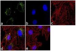
- Experimental details
- Immunofluorescence analysis of OXPHOS COMPLEX IV SU (MTCO2) was performed using 90% confluent log phase Hep G2 cells. The cells were fixed with 4% paraformaldehyde for 10 minutes, permeabilized with 0.1% Triton™ X-100 for 10 minutes, and blocked with 1% BSA for 1 hour at room temperature. The cells were labeled with MTCO2 (12C4F12) Mouse Monoclonal Antibody (Product # A-6404) at 2µg/mL in 0.1% BSA and incubated for 3 hours at room temperature and then labeled with Goat anti-Mouse IgG (H+L) Superclonal™ Secondary Antibody, Alexa Fluor® 488 conjugate (Product # A28175) at a dilution of 1:2000 for 45 minutes at room temperature (Panel a: green). Nuclei (Panel b: blue) were stained with SlowFade® Gold Antifade Mountant with DAPI (Product # S36938). F-actin (Panel c: red) was stained with Alexa Fluor® 555 Rhodamine Phalloidin (Product # R415, 1:300). Panel d represents the merged image showing membrane localization. Panel e shows the no primary antibody control. The images were captured at 60X magnification.
Supportive validation
- Submitted by
- Invitrogen Antibodies (provider)
- Main image
- Experimental details
- NULL
- Submitted by
- Invitrogen Antibodies (provider)
- Main image
- Experimental details
- NULL
- Submitted by
- Invitrogen Antibodies (provider)
- Main image

- Experimental details
- NULL
- Submitted by
- Invitrogen Antibodies (provider)
- Main image
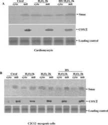
- Experimental details
- NULL
- Submitted by
- Invitrogen Antibodies (provider)
- Main image
- Experimental details
- NULL
- Submitted by
- Invitrogen Antibodies (provider)
- Main image
- Experimental details
- NULL
- Submitted by
- Invitrogen Antibodies (provider)
- Main image
- Experimental details
- NULL
- Submitted by
- Invitrogen Antibodies (provider)
- Main image
- Experimental details
- NULL
- Submitted by
- Invitrogen Antibodies (provider)
- Main image

- Experimental details
- NULL
- Submitted by
- Invitrogen Antibodies (provider)
- Main image
- Experimental details
- NULL
- Submitted by
- Invitrogen Antibodies (provider)
- Main image
- Experimental details
- NULL
- Submitted by
- Invitrogen Antibodies (provider)
- Main image
- Experimental details
- NULL
- Submitted by
- Invitrogen Antibodies (provider)
- Main image
- Experimental details
- NULL
- Submitted by
- Invitrogen Antibodies (provider)
- Main image
- Experimental details
- NULL
- Submitted by
- Invitrogen Antibodies (provider)
- Main image
- Experimental details
- NULL
- Submitted by
- Invitrogen Antibodies (provider)
- Main image
- Experimental details
- NULL
- Submitted by
- Invitrogen Antibodies (provider)
- Main image
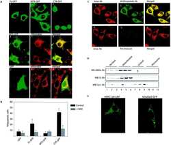
- Experimental details
- NULL
- Submitted by
- Invitrogen Antibodies (provider)
- Main image
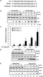
- Experimental details
- NULL
- Submitted by
- Invitrogen Antibodies (provider)
- Main image
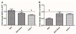
- Experimental details
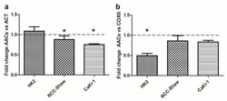
- Figure 3 Ratio of AACs protein content estimated through WB in HK2, RCC-Shaw, and CaKi-1 cells grown in serum-free medium versus complete medium conditions with respect to actin content (panel a ) or COXII content (panel b ). Data are presented as mean + SE of at least three independent experiments. * p < 0.05; nonparametric Wilcoxon two-tailed test between starved and physiological conditions. For a representative WB see Supplementary Figure S3 . The protein concentration from extraction assays was reported in Supplementary Table S2 .
- Submitted by
- Invitrogen Antibodies (provider)
- Main image
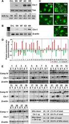
- Experimental details
- NULL
- Submitted by
- Invitrogen Antibodies (provider)
- Main image
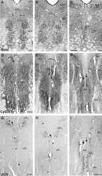
- Experimental details
- NULL
- Submitted by
- Invitrogen Antibodies (provider)
- Main image
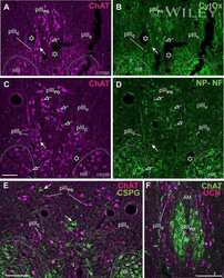
- Experimental details
- NULL
- Submitted by
- Invitrogen Antibodies (provider)
- Main image
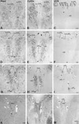
- Experimental details
- NULL
- Submitted by
- Invitrogen Antibodies (provider)
- Main image
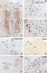
- Experimental details
- NULL
- Submitted by
- Invitrogen Antibodies (provider)
- Main image
- Experimental details
- NULL
- Submitted by
- Invitrogen Antibodies (provider)
- Main image
- Experimental details
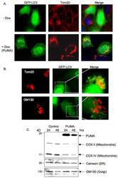
- Figure 2 PUMA induces accumulation of GFP-LC3 puncta, which preferentially colocalize with mitochondria. A) Saos-2 TetOn PUMA cells were infected with GFP-LC3 expressing Adenovirus, left for 16 hours and then treated with doxycycline. Cells were maintained in the presence of z-VAD-fmk throughout the experiment. 24 hours later, cells were fixed and immunostained with anti-Tom20 antibody to visualize the mitochondria. B) U2OS cells were infected with GFP-LC3 expressing Adenovirus, left for 16 hours and then transfected with PUMA expression plasmid. Cells were maintained in the presence of z-VAD-fmk throughout the experiment. 24 hours later, cells were fixed and immunostained with anti-Tom20 (upper panel) or anti-GM130 (lower panel). Cells exhibiting GFP-LC3 puncta were then observed under a fluorescence microscope to compare the co-localization of GFP-LC3 puncta with mitchondria (Tom20) or Golgi (GM130). C) U2OS cells were transfected with vector control or PUMA expression plasmid and maintained in the presence of z-VAD-fmk throughout the experiment. Cells were harvested 24 or 48 hours after transfection and cell lysates were immunoblotted with the indicated antibodies to detect levels of mitochondria, Golgi and ER proteins.
- Submitted by
- Invitrogen Antibodies (provider)
- Main image
- Experimental details
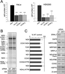
- Figure 4 Depletion of ERAL1 leads to apoptosis prior to appreciable loss of mitochondrial protein synthesis ( A ) Counts of HEK-293T or HeLa cells were taken after siRNA treatment (3 or 4 days respectively) with an NT control or each of the three independent siRNAs targeted either to the EraL1 open reading frame (ORF1; ORF2) or to the corresponding 3'-UTR. Counts were performed on three (HeLa) or six (HEK-293T) independent repeats. *** P
- Submitted by
- Invitrogen Antibodies (provider)
- Main image
- Experimental details
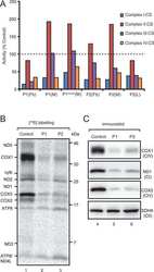
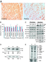
- Fig. 3 Effect of FARS2 mutation on mitochondrial homeostasis. A. Cytochrome c oxidase (COX) histochemistry of the patient muscle showed a generalised loss of enzyme activity compared to age-matched control tissue. B. Respiratory chain enzyme activity in muscle biopsy, fibroblast and myoblast: activities of complex I, complex II, and complex IV were determined in control (blue) and patient (red) and normalised to citrate synthase. Results are based on three independent measurements and are shown as percent of the mean control value +- standard deviation C. Steady state levels of RC proteins in fibroblasts (left panel) and myoblasts (right panel) were determined by Western blotting. 10% SDS-PAGE was performed with cell lysates (30 mug) from control (C1, C2) and patient (P), except for FARS2 and SDHA in the bottom 2 panels where 80 mug mitochondrial protein was loaded per lane. Western blots were decorated with antibodies to the proteins indicated. Secondary alpha-antibodies were HRP conjugated and detection was by ECL + and ImageQuant software. D. High resolution northern blot analysis was performed on total RNA (2 mug) from control (C1, C2) and patient (P). Membranes were hybridised with radiolabelled probes for mt-tRNA Phe , mt-tRNA Val and mt-tRNA Leu(UUR) . Densitometric analyses were performed on all blots. A representative example is presented with the values relative to controls for the signals derived for each tRNA below the sample. E. De novo mitochondrial protein synt
- Submitted by
- Invitrogen Antibodies (provider)
- Main image
- Experimental details
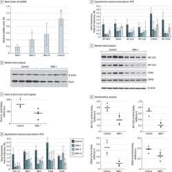
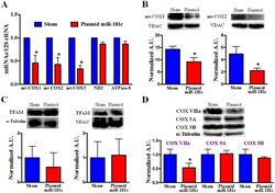
- Figure 5 Mitochondrial Complex IV Remodeling after miR-181c Treatment. (A) qPCR data show that overexpression of miR-181c significantly reduces the mRNA levels of all mitochondrial complex IV genes with 3 weeks treatment. The treatment protocol has no effect on other mitochondrial genes, such as ND2 (complex I) and ATPase 8 (complex V). Content of mRNA was first normalized to 12S rRNA, a mitochondrial gene, as 12S rRNA expression did not change with miR-181c overexpresssion. Then we normalized the data to the sham group. *p
- Submitted by
- Invitrogen Antibodies (provider)
- Main image
- Experimental details
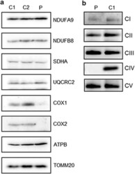
- Figure 2 Steady-state levels of OXPHOS components and complexes. ( a ) Cell lysate from control (C1 and C2) and patient (P) fibroblasts (40 mu g) were analysed by SDS-PAGE (12%) and immunoblotting. Subunit-specific antibodies were used against CI (NDUFA9, NDUFB8), CII (SDHA), CIII (UQCRC2), CIV (COX1, COX2) and CV (ATPB). The outer mitochondrial membrane marker, TOM20, was used as a loading control. ( b ) Mitochondrial proteins (50 mu g) isolated from patient (P) and control (C1) fibroblasts were analysed by one-dimensional BN-PAGE (4 to 16% gradient) using subunit-specific antibodies as indicated (CI (NDUFA9), CII (SDHA), CIII (UQCRC2), CIV (COX1) and CV (ATP5A)) to assess the assembly of individual OXPHOS complexes. Complex II (SDHA) was used as a loading control.
- Submitted by
- Invitrogen Antibodies (provider)
- Main image
- Experimental details
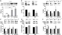
- Fig. 2. Assessment of mitochondrial bioenergetics in neuronal tissues (brainstem, posterior columns, nerve roots and dorsal root ganglia). A quantitative western blot assay was developed to measure FXN (A), COXI and COXII (B), cytochrome c (C), ATP synthase (D) and TOM22 (E) in neuronal tissues [brainstem, posterior columns (PC), nerve roots and dorsal root ganglia (DRG)]. (A) Representative western blot of human FXN expression in YG8R mice in the four neuronal tissues used in the study. Human FXN was only detected in YG8R mice, in which the transgene was present, and not in wild type (WT; data not shown). Western blot results were quantified for each lane using Fujifilm's Multi-Gauge software. To allow for loading variation, values were normalized to the actin control. Final values were expressed as a ratio to the value of FXN expression in nerve roots. DRG and nerve roots (distal tissues) expressed less FXN than the posterior columns and brainstem (proximal tissues). Values are expressed as mean+-s.e.m.; *** P
- Submitted by
- Invitrogen Antibodies (provider)
- Main image
- Experimental details
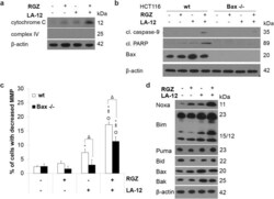
- Fig 2 Involvement of mitochondria in cooperative cytotoxic action of rosiglitazone and LA-12. (a) The release of cytochrome c into the cytoplasm (cytoplasmic fraction) of HCT116 cells pretreated (24 h) with rosiglitazone (RGZ, 50 muM) and subsequently treated (48 h) with LA-12 (0.75 muM), detected by Western blotting after cell fractionation. (b) Cleavage of caspase-9, PARP, Bax protein level (Western blotting), and (c) changes in mitochondrial membrane potential (MMP, flow cytometry) in HCT116 wt and Bax-/- cells treated as in a). (d) The level of Noxa, Bim, Puma, Bid, Bax and Bak protein (Western blotting) in HCT116 wt cells treated as in a). Results are means + S.E.M. or representatives of three independent experiments. Statistical significance: P < 0.05, * versus control, ++ versus RGZ or Omicron versus LA-12, and Delta for wt versus Bax-/- cells.
- Submitted by
- Invitrogen Antibodies (provider)
- Main image
- Experimental details
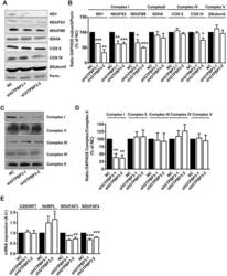
- Fig 7 Stable knock-down of GTPBP3 disturbs Complex I assembly and reduces the expression of Complex I assembly factors NDUFAF3 and NDUFAF4 (A) Western blot analysis of OXPHOS subunits ND1, NDUFS3 and NDUFB8 (Complex I), SDHA (Complex II), COXII and COXIV (Complex IV), and beta-subunit (Complex V) in shGTPBP3-1, shGTPBP3-2 and negative control (NC) cells. The filter was also probed with porin as a loading control. (B) Densitometric analysis of OXPHOS subunits normalized to porin and represented as % of NC. (C) Representative Blue Native-PAGE of OXPHOS complexes in shGTPBP3-1, shGTPBP3-2 and NC cells. (D) Densitometric analysis of OXPHOS Complexes normalized to Complex-II (loading control) and represented as % of NC. (E) qRT-PCR analysis of C20ORF7 , NUBPL , NDUFAF3 and NDUFAF4 mRNA expression in shGTPBP3 and NC cells. All data are the mean +- SEM of at least three independent biological replicates. Differences from NC values were found to be statistically significant at *p
- Submitted by
- Invitrogen Antibodies (provider)
- Main image
- Experimental details
- Figure 3 Ratio of AACs protein content estimated through WB in HK2, RCC-Shaw, and CaKi-1 cells grown in serum-free medium versus complete medium conditions with respect to actin content (panel a ) or COXII content (panel b ). Data are presented as mean + SE of at least three independent experiments. * p < 0.05; nonparametric Wilcoxon two-tailed test between starved and physiological conditions. For a representative WB see Supplementary Figure S3 . The protein concentration from extraction assays was reported in Supplementary Table S2 .
- Submitted by
- Invitrogen Antibodies (provider)
- Main image
- Experimental details
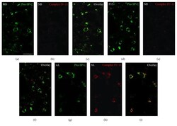
- Figure 8 Representative immunofluorescence analysis of mitochondrial complex IV subunit II protein in AECII from lung tissue sections of newborn (NB), P15, and adult (AL) animals. Lung tissue samples from the three postnatal stages were embedded into paraffin. Thereafter, 3 mu m paraffin sections were cut with a rotation microtome and processed further for indirect double immunofluorescence.The lung sections were incubated overnight for double labelling with primary antibodies against mitochondrial complex IV subunit II and pro-SP-C, a marker for AECII ( Table 1 ). The following morning, the sections were washed and incubated with the secondary antibodies ( Table 1 ) for 2 h at room temperature. Double fluorescence samples were analyzed by confocal laser scanning microscopy (CLSM) with a Leica TCS SP5. (a, d, and g) Double immunofluorescence stainings of AECII with their marker protein pro-SP-C. (b, e, and h) IF preparations for the mitochondrial complex IV subunit II. (c, f, and i) Double IF overlay for complex IV subunit II combined with pro-SP-C. NB, newborn; P15, postnatal day 15; AL, adult. Bars represent 20 mu m.
- Submitted by
- Invitrogen Antibodies (provider)
- Main image
- Experimental details
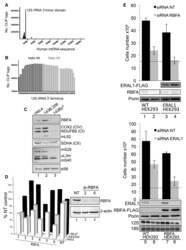
- Figure 2. RBFA binds to helix 45 of 12S rRNA but has a distinct function from that of ERAL1. ( A ) The graph represents the human mtDNA sequence with locations and numbers of the CLIP tags indicated. Tags were generated as described in the Experimental Procedures. ( B ) The location of the greatest number of CLIP tags from three independent experiments is depicted spanning helix 45 and protruding into helix 44. The two dimethylated adenines in helix 45 are indicated in light grey. The terminal C-residue represents the near 3'-terminus of the 12S mt-rRNA, corresponding to nt1597 of mtDNA. ( C ) Lysates (30 mug) from HeLa, 143B.206 parental and Rho 0 cells were analyzed by western blot to compare the relative expression levels of RBFA, components of the OXPHOS complexes (COX2, NDUFB8 and SDHA) and members of the mitoribosome (mS29, mS40, mL62 and uL3m,). Cytosolic RP-S6 is also shown. ( D ) Cell growth of each cell line was determined after 3 days treatment of six RBFA-targeted siRNAs (33 nM) (lanes 1-6) compared with control (NT lane 7). Inset: western blot of cell lysates (25 ug) after treatment with RBFA siRNA 2 and 6 to assess the level of depletion with beta-actin as the loading control. ( E ) Top panel: HEK293 cells were grown for 72 h in the presence of either si-NT (lanes 1 and 3) or si-RBFA (lanes 2 and 4). ERAL1-FLAG expression was induced 4 h after siRNA transfection (lanes 3 and 4). Lower panel: HEK293 cells were grown for 72 h in the presence of either si-NT (lane
- Submitted by
- Invitrogen Antibodies (provider)
- Main image
- Experimental details
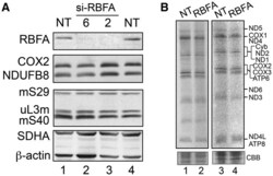
- Figure 3. Depletion of RBFA has no appreciable effect on mitochondrial protein synthesis. ( A ) HEK293 cells were treated with RBFA (lanes 2 and 3) or non-targeting (NT, lanes 1 and 4) siRNA for 3 days and extracts (25 ug) were subjected to western blotting to compare OXPHOS (COX2, NDUFB8 and SDHA) or mitoribosomal (mS40, mS29 and uL3m) protein levels. ( B ) Following similar siRNA treatment (NT or RBFA-2) for 3 (lanes 1 and 2) or 6 (lanes 3 and 4) days, cells were metabolically labelled ( 35 S-met/cys) and extracts (3 days--30 ug; 6 days--50 ug) separated by 15% denaturing PAGE. Migration of the 13 mtDNA-encoded polypeptides is indicated and loading confirmed by Coomassie blue (CBB) staining.
- Submitted by
- Invitrogen Antibodies (provider)
- Main image
- Experimental details
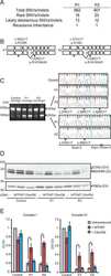
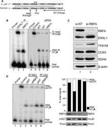
- Figure 4. Depletion of RBFA causes a decrease in 12S rRNA modification. ( A ) Schematic presenting sequence conservation of helix 45 (dashed line) and the primer extension assay used to measure the modification levels at adenine residues A 936 / A 937 in human 12S rRNA. If the modification is present, the primer extends four residues, and in its absence the primer extends a further five residues. ( B ) To determine the modification status of 12S rRNA bound to ERAL1-FLAG (lane 2, 3-day induction) or RBFA-FLAG (lane 3, 3 days), these proteins were immunoprecipitated from HEK293 cells and the bound RNA was extracted. Samples were subjected to primer extension and denaturing PAGE. (Right panel) Cells were depleted of ERAL1 (3 days) followed by 4 days of siRNA treatment; si-NT (lane 7) or si-RBFA (lane 8). RNA was extracted and primer extension performed. Primer alone (lane 1, 4), extension on unmethylated template (lane 5) and wild-type cells (lane 6) were controls performed in parallel. ( C ) Western blot (50 ug of cell extracts) determined the steady-state levels of ERAL1, TFB1M and COXII in cells depleted of RBFA. SDHA and beta-actin were used as loading controls. ( D ) HEK293 cells were grown for 5 days in the presence of NT or RBFA siRNA with induction of mS27-FLAG (IP SSU) or mL62-FLAG (IP LSU) on day 3. (Left panel) Cell extracts were subjected to immunoprecipitation and bound 12S rRNA was assessed by primer extension alongside controls (primer alone, lane 1; unmethylate
 Explore
 Validate
 Learn
Learn Western blot
 Immunocytochemistry