Explore
 Validate
 Learn
Learn Western blot
Antibody data
- Antibody Data
- Antigen structure
- References [3]
- Comments [0]
- Validations
- Western blot [5]
- Other assay [3]
Submit
Validation data
Reference
Comment
Report error
- Product number
- 702785 - Provider product page

- Provider
- Invitrogen Antibodies
- Product name
- Parkin Recombinant Rabbit Monoclonal Antibody (21H24L9)
- Antibody type
- Monoclonal
- Antigen
- Other
- Description
- This antibody is predicted to react with Monkey, Rat, Pig, Mouse.
- Antibody clone number
- 21H24L9
- Concentration
- 0.5 mg/mL
Submitted references Mitochondrial-Targeted Therapies Require Mitophagy to Prevent Oxidative Stress Induced by SOD2 Inactivation in Hypertrophied Cardiomyocytes.
LncRNA SNHG17 knockdown promotes Parkin-dependent mitophagy and reduces apoptosis of podocytes through Mst1.
Functional Pathway Identification With CRISPR/Cas9 Genome-wide Gene Disruption in Human Dopaminergic Neuronal Cells Following Chronic Treatment With Dieldrin.
Peugnet V, Chwastyniak M, Mulder P, Lancel S, Bultot L, Fourny N, Renguet E, Bugger H, Beseme O, Loyens A, Heyse W, Richard V, Amouyel P, Bertrand L, Pinet F, Dubois-Deruy E
Antioxidants (Basel, Switzerland) 2022 Apr 6;11(4)
Antioxidants (Basel, Switzerland) 2022 Apr 6;11(4)
LncRNA SNHG17 knockdown promotes Parkin-dependent mitophagy and reduces apoptosis of podocytes through Mst1.
Guo F, Wang W, Song Y, Wu L, Wang J, Zhao Y, Ma X, Ji H, Liu Y, Li Z, Qin G
Cell cycle (Georgetown, Tex.) 2020 Aug;19(16):1997-2006
Cell cycle (Georgetown, Tex.) 2020 Aug;19(16):1997-2006
Functional Pathway Identification With CRISPR/Cas9 Genome-wide Gene Disruption in Human Dopaminergic Neuronal Cells Following Chronic Treatment With Dieldrin.
Russo M, Sobh A, Zhang P, Loguinov A, Tagmount A, Vulpe CD, Liu B
Toxicological sciences : an official journal of the Society of Toxicology 2020 Aug 1;176(2):366-381
Toxicological sciences : an official journal of the Society of Toxicology 2020 Aug 1;176(2):366-381
No comments: Submit comment
Supportive validation
- Submitted by
- Invitrogen Antibodies (provider)
- Main image

- Experimental details
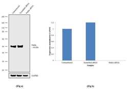
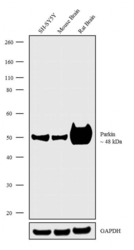
- Western blot analysis was performed on Whole cell extracts (60 µg lysate) of SH-SY5Y (Lane 1) and tissue extracts of Mouse Brain (Lane 2) and Rat Brain (Lane 3). The blots were probed with Anti-Parkin Recombinant Rabbit Monoclonal Antibody (Product # 702785, 2.5 µg/mL) and detected by chemiluminescence using Goat anti-Rabbit IgG (H+L) Superclonal™ Secondary Antibody, HRP conjugate (Product # A27036, 0.25 µg/mL, 1:4000 dilution). A 48 kDa band corresponding to Parkin was observed across the cell line and tissues tested. Known quantity of protein samples were electrophoresed using Novex®NuPAGE®4-12% Bis-Tris gel (Product # NP0322BOX), XCell SureLock™ Electrophoresis System (Product # EI0002) and Novex® Sharp Pre-Stained Protein Standard (Product # LC5800). Resolved proteins were then transferred onto a nitrocellulose membrane with iBlot® Dry Blotting System (Product # IB21001). The membrane was probed with the relevant primary and secondary Antibody following blocking with 5% skimmed milk. Chemiluminescent detection was performed using Pierce™ ECL Western blotting Substrate (Product # 32106).

- Submitted by
- Invitrogen Antibodies (provider)
- Main image
- Experimental details
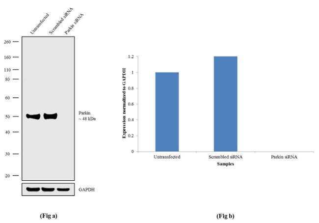
- Knockdown of Parkin was achieved by transfecting SH-SY5Y cells with Parkin specific siRNA (Silencer® select Product # s10043+s10045). Western blot analysis (Fig a) was performed using whole cell extracts from Parkin knockdown cells (Lane 3), non-specific scrambled siRNA transfected cells (Lane 2) and untransfected cells (Lane 1). The blots were probed with Anti-Parkin Recombinant Rabbit Monoclonal Antibody (Product # 702785, 1-3 µg/mL) and Goat anti-Rabbit IgG (H+L) Superclonal™ Secondary Antibody, HRP conjugate (Product # A27036, 0.25 µg/mL, 1:4000 dilution). Densitometric analysis of this Western blot is shown in histogram (Fig b). Loss of signal upon siRNA mediated knock down confirms that antibody is specific to Parkin.
- Submitted by
- Invitrogen Antibodies (provider)
- Main image
- Experimental details
- Western blot analysis was performed on Whole cell extracts (60 µg lysate) of SH-SY5Y (Lane 1) and tissue extracts of Mouse Brain (Lane 2) and Rat Brain (Lane 3). The blots were probed with Anti-Parkin Recombinant Rabbit Monoclonal Antibody (Product # 702785, 2.5 µg/mL) and detected by chemiluminescence using Goat anti-Rabbit IgG (H+L) Superclonal™ Secondary Antibody, HRP conjugate (Product # A27036, 0.25 µg/mL, 1:4000 dilution). A 48 kDa band corresponding to Parkin was observed across the cell line and tissues tested. Known quantity of protein samples were electrophoresed using Novex®NuPAGE®4-12% Bis-Tris gel (Product # NP0322BOX), XCell SureLock™ Electrophoresis System (Product # EI0002) and Novex® Sharp Pre-Stained Protein Standard (Product # LC5800). Resolved proteins were then transferred onto a nitrocellulose membrane with iBlot® Dry Blotting System (Product # IB21001). The membrane was probed with the relevant primary and secondary Antibody following blocking with 5% skimmed milk. Chemiluminescent detection was performed using Pierce™ ECL Western blotting Substrate (Product # 32106).

- Submitted by
- Invitrogen Antibodies (provider)
- Main image
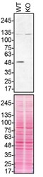
- Experimental details
- Western blot of Parkin was performed by loading 50 µg of WT (lane 1) and PRKN CRISPR KO (lane 2) SH-SY5Y cell lysates in RIPA buffer onto a 4-15% gradient polyacrylamide gel. Proteins on the blots were visualized with Ponceau staining (below immunoblot). Proteins were transferred to nitrocellulose membrane and blocked in 5% milk for 1 hr. PRKN was detected at approximately 52 kDa using a PRKN recombinant monoclonal antibody (Product # 702785) at a dilution of 1:200 in 5% BSA in TBS with 0.1% Tween 20 (TBST) overnight at 4°C. The peroxidase-conjugated secondary antibody (Product # 65-6120) was diluted to 0.2 µg/mL in TBST with 5% milk for 1 hr. Chemiluminescent detection was performed using Pierce ECL Western Blotting Substrate (Product # 32106). Data courtesy of YCharOS Inc., an open science company with the mission of characterizing commercially available antibodies using knockout validation.

- Submitted by
- Invitrogen Antibodies (provider)
- Main image
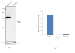
- Experimental details
- Western blot analysis (Fig a) of Parkin was performed on cell extracts (100 µg of lysate) of HEK-293 wild type (Lane 1) and Parkin knockout (Lane 2). The blot was probed with Anti-Parkin Recombinant Rabbit Monoclonal Antibody (Product # 702785, 1:500 dilution) and detected by chemiluminescence using Peroxidase AffiniPure Goat anti-Rabbit IgG (H+L) Secondary Antibody, HRP conjugate (Product # 111-035-144, 1:4000 dilution). Densitometric analysis of this Western blot is shown in the histogram (Fig b). Loss of signal upon CRISPR mediated knockout (KO) confirms that antibody is specific to Parkin.

Supportive validation
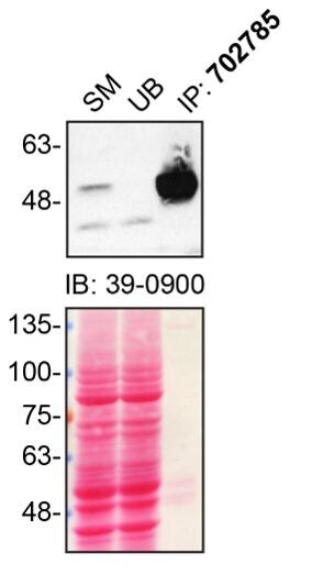
- Submitted by
- Invitrogen Antibodies (provider)
- Main image
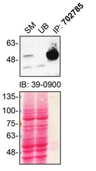
- Experimental details
- Immunoprecipitation of PRKN was performed on SH-SY5Y cell lysates. Antibody-bead conjugates were prepared by adding 1 µg of PRKN recombinant monoclonal antibody (Product # 702785) with 30 µL of protein A-Sepharose beads and rocked overnight at 4°C. 2 mg of lysate was incubated with an antibody-bead conjugate for 2 hours at 4°C. Following centrifugation and multiple washes, 10% starting material (SM), 10% unbound fraction (UB) and immunoprecipitated fraction (IP) were processed for immunoblot using another PRKN monoclonal antibody (Product # 39-0900). Ponceau stained transfer of blot is shown. Data courtesy of YCharOS Inc., an open science company with the mission of characterizing commercially available antibodies using knockout validation.
- Submitted by
- Invitrogen Antibodies (provider)
- Main image
- Experimental details
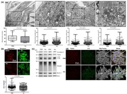
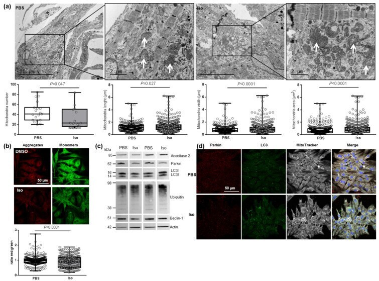
- Characterization of mitochondrial function and autophagy/mitophagy in hypertrophied neonatal rat cardiomyocytes (NCMs). ( a ) Ultrastructure of PBS- and Iso-treated NCMs (magnification x7000 with a scale bar of 10 um (left images) and x12,000 with a scale bar of 2 um (right images)) and quantification of mitochondria number, length (um), width (um) and area (um 2 ). Arrows indicate example of mitochondria for comparison. ( b ) Mitochondrial membrane potential was quantified in NCMs treated with Iso by fluorescence quantification of JC-1 dye for aggregates (red) and monomer (green) (from 3 independent experiments and at least 366 cells). ( c ) Representative images for quantification of Krebs cycle by Western blot of aconitase 2 and mitophagy/autophagy by Western blot of parkin, LC3II/LC3I ratio, ubiquitin and beclin-1 in NCMs treated with Iso Data were normalized to actin. ( d ) Representative images of parkin (red) and LC3 (green) localized in mitochondria (white) of untreated (PBS) and Iso-treated NCMs. Colocalization appeared in merge images. Nuclei were stained by Dapi (blue). Only significant p values are indicated from at least 3 independent experiments. Images were selected to represent the mean values of each condition.
- Submitted by
- Invitrogen Antibodies (provider)
- Main image
- Experimental details
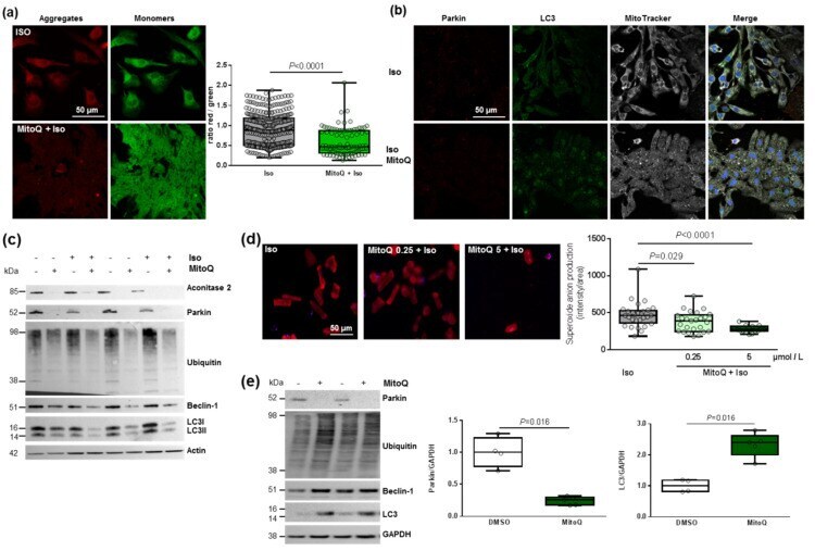
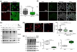
- Effect of MitoQ on mitophagy in cardiomyocytes. ( a ) Mitochondrial membrane potential was quantified in hypertrophied NCM with pre-treatment with mitoquinone (MitoQ) by fluorescence quantification of JC-1 dye for aggregates (red) and monomer (green) (from 3 independent experiments and at least 94 cells). ( b ) Representative images of parkin (red) and LC3 (green) localized in mitochondria (white) of Iso-treated NCMs with pre-treatment with mitoquinone (MitoQ). Colocalization appeared in merge images. Nuclei were stained by Dapi (blue). ( c ) Mitophagy was quantified in control (-) or Iso-treated NCMs for 24 h (+) with (+) or without (-) MitoQ by Western blot of parkin, ubiquitinated proteins, beclin-1 and LC3II/LC3I ratio. Data were normalized to actin. ( d ) Mitochondrial superoxide anion was quantified in adult cardiomyocytes (ACMs) treated with Iso for 48 h with or without mitoQ pre-treatment (0.25 and 5 umol/L) by fluorescence quantification of mitoSOX (red) (from at least 12 cells). ( e ) Mitophagy was quantified in human cardiomyocytes (HCMs) with (+) or without (-) MitoQ pre-treatment by Western blot of parkin, ubiquitinated proteins, beclin-1 and LC3. Data were normalized to GAPDH. Only significant p values are indicated from at least 3 independent experiments. Images were selected to represent the mean values of each condition.