Explore
 Validate
 Learn
Learn Western blot
 Immunoprecipitation
Antibody data
- Antibody Data
- Antigen structure
- References [3]
- Comments [0]
- Validations
- Immunoprecipitation [1]
- Other assay [3]
Submit
Validation data
Reference
Comment
Report error
- Product number
- 702785 - Provider product page

- Provider
- Invitrogen Antibodies
- Product name
- Parkin Recombinant Rabbit Monoclonal Antibody (21H24L9)
- Antibody type
- Monoclonal
- Antigen
- Other
- Description
- This antibody is predicted to react with Monkey, Rat, Pig, Mouse. Recombinant rabbit monoclonal antibodies are produced using in vitro expression systems. The expression systems are developed by cloning in the specific antibody DNA sequences from immunoreactive rabbits. Then, individual clones are screened to select the best candidates for production. The advantages of using recombinant rabbit monoclonal antibodies include: better specificity and sensitivity, lot-to-lot consistency, animal origin-free formulations, and broader immunoreactivity to diverse targets due to larger rabbit immune repertoire.
- Reactivity
- Human, Mouse, Rat
- Host
- Rabbit
- Isotype
- IgG
- Antibody clone number
- 21H24L9
- Vial size
- 100 μg
- Concentration
- 0.5 mg/mL
- Storage
- Store at 4°C short term. For long term storage, store at -20°C, avoiding freeze/thaw cycles.
Submitted references Mitochondrial-Targeted Therapies Require Mitophagy to Prevent Oxidative Stress Induced by SOD2 Inactivation in Hypertrophied Cardiomyocytes.
LncRNA SNHG17 knockdown promotes Parkin-dependent mitophagy and reduces apoptosis of podocytes through Mst1.
Functional Pathway Identification With CRISPR/Cas9 Genome-wide Gene Disruption in Human Dopaminergic Neuronal Cells Following Chronic Treatment With Dieldrin.
Peugnet V, Chwastyniak M, Mulder P, Lancel S, Bultot L, Fourny N, Renguet E, Bugger H, Beseme O, Loyens A, Heyse W, Richard V, Amouyel P, Bertrand L, Pinet F, Dubois-Deruy E
Antioxidants (Basel, Switzerland) 2022 Apr 6;11(4)
Antioxidants (Basel, Switzerland) 2022 Apr 6;11(4)
LncRNA SNHG17 knockdown promotes Parkin-dependent mitophagy and reduces apoptosis of podocytes through Mst1.
Guo F, Wang W, Song Y, Wu L, Wang J, Zhao Y, Ma X, Ji H, Liu Y, Li Z, Qin G
Cell cycle (Georgetown, Tex.) 2020 Aug;19(16):1997-2006
Cell cycle (Georgetown, Tex.) 2020 Aug;19(16):1997-2006
Functional Pathway Identification With CRISPR/Cas9 Genome-wide Gene Disruption in Human Dopaminergic Neuronal Cells Following Chronic Treatment With Dieldrin.
Russo M, Sobh A, Zhang P, Loguinov A, Tagmount A, Vulpe CD, Liu B
Toxicological sciences : an official journal of the Society of Toxicology 2020 Aug 1;176(2):366-381
Toxicological sciences : an official journal of the Society of Toxicology 2020 Aug 1;176(2):366-381
No comments: Submit comment
Supportive validation
- Submitted by
- Invitrogen Antibodies (provider)
- Main image

- Experimental details
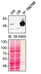
- Immunoprecipitation of PRKN was performed on SH-SY5Y cell lysates. Antibody-bead conjugates were prepared by adding 1 µg of PRKN recombinant monoclonal antibody (Product # 702785) with 30 µL of protein A-Sepharose beads and rocked overnight at 4°C. 2 mg of lysate was incubated with an antibody-bead conjugate for 2 hours at 4°C. Following centrifugation and multiple washes, 10% starting material (SM), 10% unbound fraction (UB) and immunoprecipitated fraction (IP) were processed for immunoblot using another PRKN monoclonal antibody (Product # 39-0900). Ponceau stained transfer of blot is shown. Data courtesy of YCharOS Inc., an open science company with the mission of characterizing commercially available antibodies using knockout validation.

Supportive validation
- Submitted by
- Invitrogen Antibodies (provider)
- Main image
- Experimental details
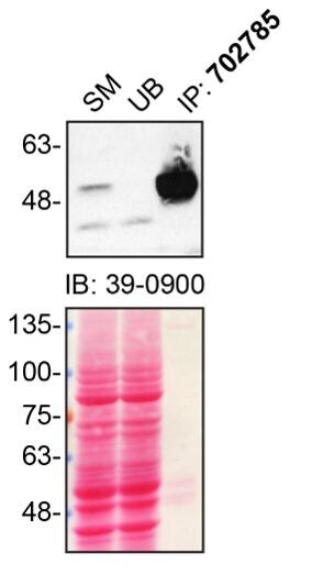
- Immunoprecipitation of PRKN was performed on SH-SY5Y cell lysates. Antibody-bead conjugates were prepared by adding 1 µg of PRKN recombinant monoclonal antibody (Product # 702785) with 30 µL of protein A-Sepharose beads and rocked overnight at 4°C. 2 mg of lysate was incubated with an antibody-bead conjugate for 2 hours at 4°C. Following centrifugation and multiple washes, 10% starting material (SM), 10% unbound fraction (UB) and immunoprecipitated fraction (IP) were processed for immunoblot using another PRKN monoclonal antibody (Product # 39-0900). Ponceau stained transfer of blot is shown. Data courtesy of YCharOS Inc., an open science company with the mission of characterizing commercially available antibodies using knockout validation.
- Submitted by
- Invitrogen Antibodies (provider)
- Main image
- Experimental details
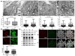
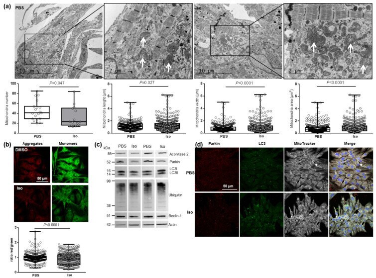
- Characterization of mitochondrial function and autophagy/mitophagy in hypertrophied neonatal rat cardiomyocytes (NCMs). ( a ) Ultrastructure of PBS- and Iso-treated NCMs (magnification x7000 with a scale bar of 10 um (left images) and x12,000 with a scale bar of 2 um (right images)) and quantification of mitochondria number, length (um), width (um) and area (um 2 ). Arrows indicate example of mitochondria for comparison. ( b ) Mitochondrial membrane potential was quantified in NCMs treated with Iso by fluorescence quantification of JC-1 dye for aggregates (red) and monomer (green) (from 3 independent experiments and at least 366 cells). ( c ) Representative images for quantification of Krebs cycle by Western blot of aconitase 2 and mitophagy/autophagy by Western blot of parkin, LC3II/LC3I ratio, ubiquitin and beclin-1 in NCMs treated with Iso Data were normalized to actin. ( d ) Representative images of parkin (red) and LC3 (green) localized in mitochondria (white) of untreated (PBS) and Iso-treated NCMs. Colocalization appeared in merge images. Nuclei were stained by Dapi (blue). Only significant p values are indicated from at least 3 independent experiments. Images were selected to represent the mean values of each condition.
- Submitted by
- Invitrogen Antibodies (provider)
- Main image
- Experimental details
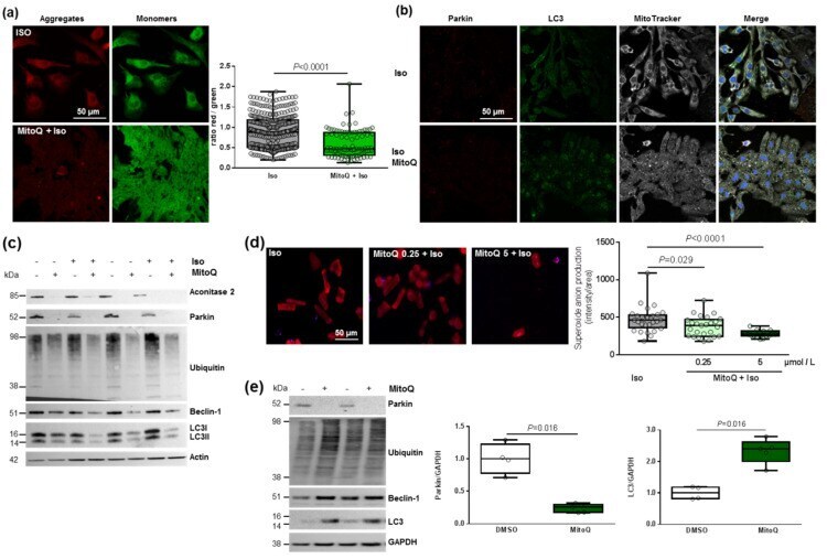
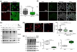
- Effect of MitoQ on mitophagy in cardiomyocytes. ( a ) Mitochondrial membrane potential was quantified in hypertrophied NCM with pre-treatment with mitoquinone (MitoQ) by fluorescence quantification of JC-1 dye for aggregates (red) and monomer (green) (from 3 independent experiments and at least 94 cells). ( b ) Representative images of parkin (red) and LC3 (green) localized in mitochondria (white) of Iso-treated NCMs with pre-treatment with mitoquinone (MitoQ). Colocalization appeared in merge images. Nuclei were stained by Dapi (blue). ( c ) Mitophagy was quantified in control (-) or Iso-treated NCMs for 24 h (+) with (+) or without (-) MitoQ by Western blot of parkin, ubiquitinated proteins, beclin-1 and LC3II/LC3I ratio. Data were normalized to actin. ( d ) Mitochondrial superoxide anion was quantified in adult cardiomyocytes (ACMs) treated with Iso for 48 h with or without mitoQ pre-treatment (0.25 and 5 umol/L) by fluorescence quantification of mitoSOX (red) (from at least 12 cells). ( e ) Mitophagy was quantified in human cardiomyocytes (HCMs) with (+) or without (-) MitoQ pre-treatment by Western blot of parkin, ubiquitinated proteins, beclin-1 and LC3. Data were normalized to GAPDH. Only significant p values are indicated from at least 3 independent experiments. Images were selected to represent the mean values of each condition.