Explore
 Validate
 Learn
Learn Western blot
 Immunocytochemistry
Antibody data
- Antibody Data
- Antigen structure
- References [2]
- Comments [0]
- Validations
- Immunocytochemistry [2]
- Immunohistochemistry [2]
- Flow cytometry [2]
- Other assay [2]
Submit
Validation data
Reference
Comment
Report error
- Product number
- PA5-13399 - Provider product page

- Provider
- Invitrogen Antibodies
- Product name
- Parkin Polyclonal Antibody
- Antibody type
- Polyclonal
- Antigen
- Synthetic peptide
- Reactivity
- Human, Mouse
- Host
- Rabbit
- Isotype
- IgG
- Vial size
- 400 μL
- Concentration
- 0.5 mg/mL
- Storage
- Store at 4°C short term. For long term storage, store at -20°C, avoiding freeze/thaw cycles.
Submitted references Mutated FANCA Gene Role in the Modulation of Energy Metabolism and Mitochondrial Dynamics in Head and Neck Squamous Cell Carcinoma.
Mitochondrial fission and mitophagy are independent mechanisms regulating ischemia/reperfusion injury in primary neurons.
Bertola N, Degan P, Cappelli E, Ravera S
Cells 2022 Jul 30;11(15)
Cells 2022 Jul 30;11(15)
Mitochondrial fission and mitophagy are independent mechanisms regulating ischemia/reperfusion injury in primary neurons.
Anzell AR, Fogo GM, Gurm Z, Raghunayakula S, Wider JM, Maheras KJ, Emaus KJ, Bryson TD, Wang M, Neumar RW, Przyklenk K, Sanderson TH
Cell death & disease 2021 May 12;12(5):475
Cell death & disease 2021 May 12;12(5):475
No comments: Submit comment
Supportive validation
- Submitted by
- Invitrogen Antibodies (provider)
- Main image
- Experimental details
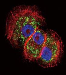
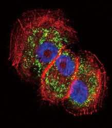
- Immunofluorescent analysis of NCI-H460 cells using a PARK2 polyclonal antibody (Product # PA5-13399) at a dilution of 1:10-50, followed by a fluor-conjugated goat anti-rabbit secondary antibody (green). Actin filaments were stained with dye-conjugated phalloidin (red). Nuclei were stained with DAPI (blue).

- Submitted by
- Invitrogen Antibodies (provider)
- Main image
- Experimental details
- Immunocytochemistry analysis of Parkin in NCI-H460 cells. Samples were incubated in Parkin polyclonal antibody (Product # PA5-13399) followed by Alexa Fluor 488-conjugated goat anti-rabbit lgG (green). Actin filaments have been labeled with Alexa Fluor 555 phalloidin (red). DAPI was used to stain the cell nuclear (blue).

Supportive validation
- Submitted by
- Invitrogen Antibodies (provider)
- Main image
- Experimental details
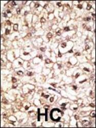
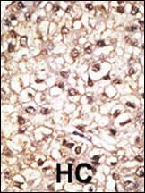
- Immunohistochemistry analysis of Parkin in formalin-fixed and paraffin-embedded human cancer tissue. Samples were incubated with Parkin polyclonal antibody (Product # PA5-13399) which was peroxidase-conjugated to the secondary antibody, followed by AEC staining. This data demonstrates the use of this antibody for immunohistochemistry; clinical relevance has not been evaluated. BC = breast carcinoma; HC = hepatocarcinoma.
- Submitted by
- Invitrogen Antibodies (provider)
- Main image
- Experimental details
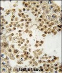
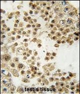
- Immunohistochemistry analysis of Parkin in formalin-fixed and paraffin-embedded human testis tissue. Samples were incubated with Parkin polyclonal antibody (Product # PA5-13399) which was peroxidase-conjugated to the secondary antibody, followed by DAB staining. This data demonstrates the use of this antibody for immunohistochemistry; clinical relevance has not been evaluated.
Supportive validation
- Submitted by
- Invitrogen Antibodies (provider)
- Main image
- Experimental details
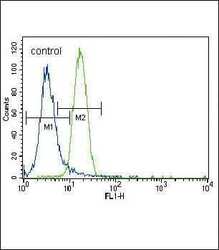
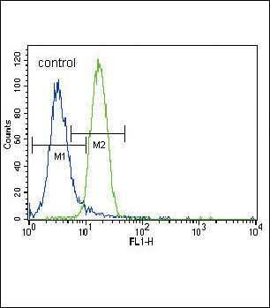
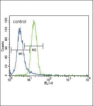
- Flow cytometry analysis of NCI-H460 cells using a PARK2 polyclonal antibody (Product # PA5-13399) (right) compared to a negative control cell (left) at a dilution of 1:10-50, followed by a FITC-conjugated goat anti-rabbit antibody
- Submitted by
- Invitrogen Antibodies (provider)
- Main image
- Experimental details
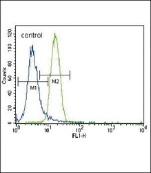
- Flow cytometry of Parkin in NCI-H460 cells (right histogram). Samples were incubated with Parkin polyclonal antibody (Product # PA5-13399) followed by FITC-conjugated goat-anti-rabbit secondary antibody. Negative control cell (left histogram).
Supportive validation
- Submitted by
- Invitrogen Antibodies (provider)
- Main image
- Experimental details
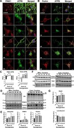
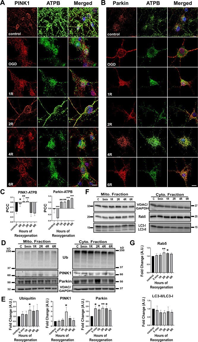
- Fig. 2 PINK1/Parkin-mediated mitophagy. A Cells labeled with PINK1 and ATPB for colocalization of PINK1 with mitochondria, n = 4. B Cells labeled with Parkin and ATPB for colocalization of Parkin with mitochondria, n = 3. C Quantification of colocalization using Pearson''s correlation coefficient (PCC). D Western Blot of mitochondrial and cytosolic fractions for PINK1, Parkin, and ubiquitin. E Quantitation of protein levels, normalized to VDAC and GAPDH, Ubiquitin: n = 6, PINK1: n = 5, Parkin: n = 8. F Western Blot of LC3 (autophagy marker) and Rab5 (endosome marker) in mitochondrial and cytosolic fractions. G Quantitation of LC3 conversion and Rab5, normalized to VDAC and GAPDH, LC3: n = 4, Rab5: n = 4. Differences between groups were computed using one-way ANOVA with Dunnett''s post-hoc analysis for comparisons versus control. R post-reoxygenation; * p < 0.05; ** p < 0.01; *** p < 0.001. Scale bar = 10 um.
- Submitted by
- Invitrogen Antibodies (provider)
- Main image
- Experimental details
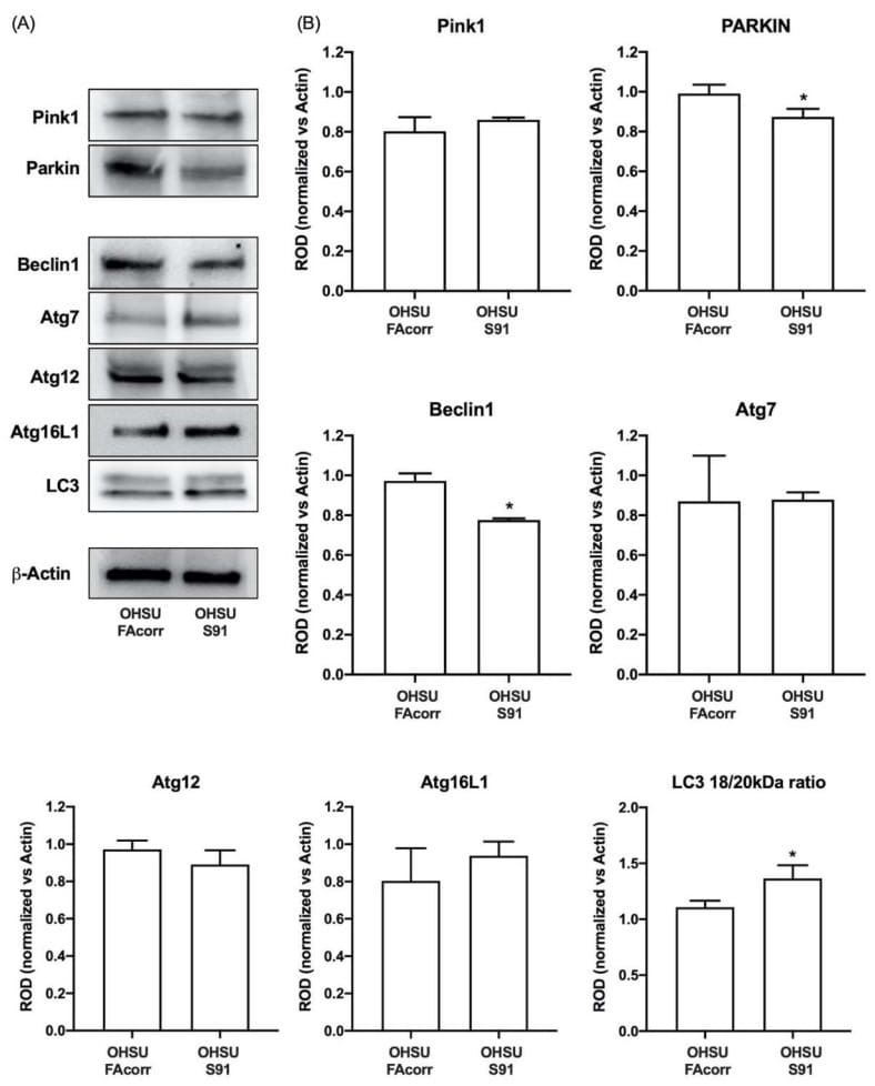
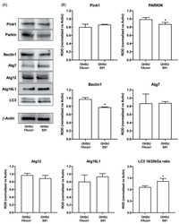
- Expression evaluation of proteins involved in mitophagy and autophagy processes in OHSU-974-FAcorr and OHSU-974-S91. ( A ) Western blot signals of Pink1 and Parkin (two mitophagy markers), Beclin1, Atg7, Atg12, Atg16L1, LC3 (autophagy markers), and beta-Actin. ( B ) Densitometric analysis of WB signals reported in Panel A, normalized versus beta-Actin. Data in histograms are reported as mean +- SD and are representative of at least 3 independent experiments. Statistical significance was tested with an unpaired t -test. * represents a p < 0.05 between OHSU-974-S91 cells and the OHSU-974-FAcorr cells used as control.