Explore
 Validate
 Learn
Learn Western blot
 Immunoprecipitation
 Other assay
Antibody data
- Antibody Data
- Antigen structure
- References [2]
- Comments [0]
- Validations
- Other assay [2]
Submit
Validation data
Reference
Comment
Report error
- Product number
- MA5-15109 - Provider product page

- Provider
- Invitrogen Antibodies
- Product name
- GSK3B Monoclonal Antibody (E.948.2)
- Antibody type
- Monoclonal
- Antigen
- Synthetic peptide
- Description
- It is not recommended to aliquot this antibody.
- Reactivity
- Human, Mouse, Rat
- Host
- Rabbit
- Isotype
- IgG
- Antibody clone number
- E.948.2
- Vial size
- 100 μL
- Concentration
- 13 μg/mL
- Storage
- -20°C
Submitted references The extracellular HDAC6 ZnF UBP domain modulates the actin network and post-translational modifications of Tau.
Melatonin Reduces GSK3β-Mediated Tau Phosphorylation, Enhances Nrf2 Nuclear Translocation and Anti-Inflammation.
Balmik AA, Sonawane SK, Chinnathambi S
Cell communication and signaling : CCS 2021 May 1;19(1):49
Cell communication and signaling : CCS 2021 May 1;19(1):49
Melatonin Reduces GSK3β-Mediated Tau Phosphorylation, Enhances Nrf2 Nuclear Translocation and Anti-Inflammation.
Das R, Balmik AA, Chinnathambi S
ASN neuro 2020 Jan-Dec;12:1759091420981204
ASN neuro 2020 Jan-Dec;12:1759091420981204
No comments: Submit comment
Supportive validation
- Submitted by
- Invitrogen Antibodies (provider)
- Main image
- Experimental details
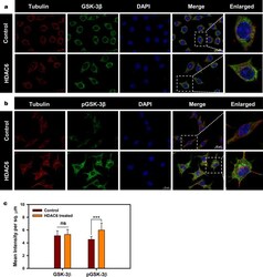
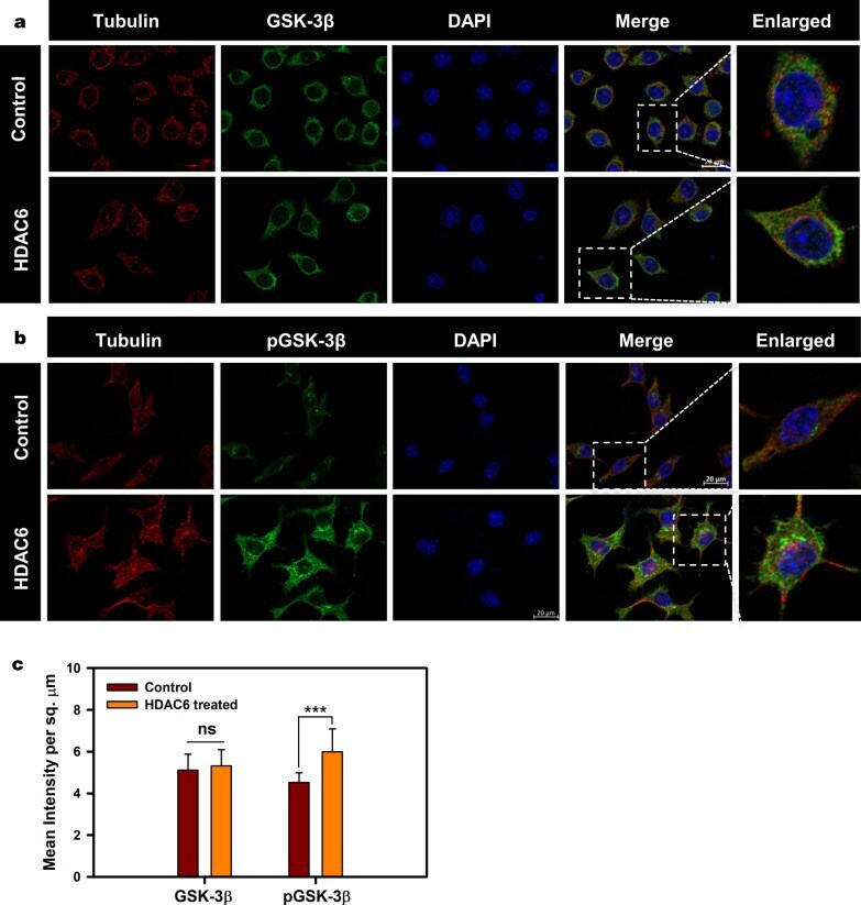
- Fig. 1 Downregulation of GSK-3beta activity by HDAC6. a Neuro2a mapped for total GSK-3beta shows their unaltered levels upon HDAC6 ZnF UBP. b Inhibitory phosphorylation of GSK-3beta at Ser9 increases upon HDAC6 ZnF UBP treatment. The enlarged image shows the elevated level of pGSK-3beta compared to neuro2a cell control. c Quantification of mean fluorescence intensity for GSK-3beta in cell control and HDAC6 ZnF UBP treated cells showed non-significant difference while the levels of pGSK-3beta were significantly increased upon HDAC6 ZnF UBP treatment in comparison to cell control
- Submitted by
- Invitrogen Antibodies (provider)
- Main image
- Experimental details
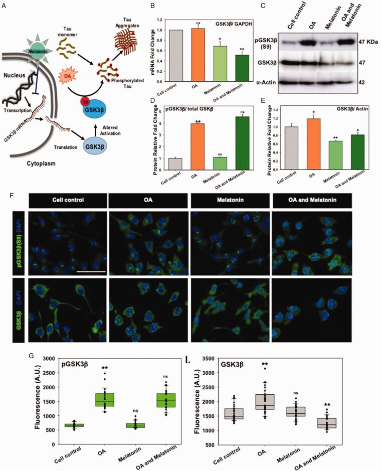
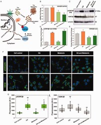
- Figure 2. Melatonin Altered GSK3beta Expression but Not Activation. A: In AD, Tau becomes hyperphosphorylated and subsequently deposits as aggregates in neurons. OA has been proven as a model to induce Tau phosphorylation via PP2A inhibition and enhancement of overall kinase activity. Melatonin has been proposed to block the GSK3beta-mRNA expression and total protein level. B: Melatonin reduced the total-GSK3beta mRNA expression level on Neuro2A cells upon OA treatment by qRT-PCR. C and D: OA has induced the Ser 9 phosphorylation of GSK3beta due to the overall enhancement of global phosphorylation, whereas the level of total GSK3beta decreased slightly. E: Western blot densitometric quantification showed a decreased level of total GSK3beta in Melatonin-treated Neuro2A cells. F: Immunofluorescence study depicted the altered level of only total GSK3beta protein, but GSK3beta (phopsho Ser9) remained invariant. G and H: The relative fluorescence level was plotted for Melatonin and OA treated group alone and together. * corresponds to p-values of test groups compared with untreated control (*p < 0.05; **p