Explore
 Validate
 Learn
Learn Western blot
 ELISA
ELISAAntibody data
- Antibody Data
- Antigen structure
- References [4]
- Comments [0]
- Validations
- Western blot [4]
- Immunohistochemistry [1]
- Flow cytometry [2]
Submit
Validation data
Reference
Comment
Report error
- Product number
- NBP1-47470 - Provider product page

- Provider
- Novus Biologicals
- Proper citation
- Novus Cat#NBP1-47470, RRID:AB_10010422
- Product name
- Mouse Monoclonal GSK-3 beta Antibody
- Antibody type
- Monoclonal
- Description
- Ammonium sulfate precipitation.
- Reactivity
- Human, Mouse, Rat, Simian
- Host
- Mouse
- Isotype
- IgG
- Vial size
- 0.1 ml
- Concentration
- 1.0 mg/ml
- Storage
- Store at 4C short term. Aliquot and store at -20C long term. Avoid freeze-thaw cycles.
Submitted references Cyclic AMP and glycogen synthase kinase 3 form a regulatory loop in spermatozoa.
Inhibition of Fas associated phosphatase 1 (Fap1) facilitates apoptosis of colon cancer stem cells and enhances the effects of oxaliplatin.
Notch signaling in response to excitotoxicity induces neurodegeneration via erroneous cell cycle reentry.
The protease Omi regulates mitochondrial biogenesis through the GSK3β/PGC-1α pathway.
Dey S, Goswami S, Eisa A, Bhattacharjee R, Brothag C, Kline D, Vijayaraghavan S
Journal of cellular physiology 2018 Sep;233(9):7239-7252
Journal of cellular physiology 2018 Sep;233(9):7239-7252
Inhibition of Fas associated phosphatase 1 (Fap1) facilitates apoptosis of colon cancer stem cells and enhances the effects of oxaliplatin.
Huang W, Bei L, Eklund EA
Oncotarget 2018 May 25;9(40):25891-25902
Oncotarget 2018 May 25;9(40):25891-25902
Notch signaling in response to excitotoxicity induces neurodegeneration via erroneous cell cycle reentry.
Marathe S, Liu S, Brai E, Kaczarowski M, Alberi L
Cell death and differentiation 2015 Nov;22(11):1775-84
Cell death and differentiation 2015 Nov;22(11):1775-84
The protease Omi regulates mitochondrial biogenesis through the GSK3β/PGC-1α pathway.
Xu R, Hu Q, Ma Q, Liu C, Wang G
Cell death & disease 2014 Aug 14;5(8):e1373
Cell death & disease 2014 Aug 14;5(8):e1373
No comments: Submit comment
Supportive validation
- Submitted by
- Novus Biologicals (provider)
- Main image

- Experimental details
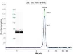
- Simple Western: GSK-3 beta Antibody (3D10) [NBP1-47470] - Simple Western lane view shows a specific band for GSK-3 Beta in 0.5 mg/ml of Hek293 lysate. This experiment was performed under reducing conditions using the 12-230 kDa separation system.

- Submitted by
- Novus Biologicals (provider)
- Main image
- Experimental details
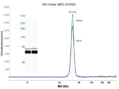
- Simple Western: GSK-3 beta Antibody (3D10) [NBP1-47470] - Simple western analysis of mouse brain tissue (striatum) from 4 month old Tg4150 and wildtype mice. Image courtesy of Dr. Brandi Wasek-Patterson at Baylor Research Institute, Institute of Metabolic Disease.
- Submitted by
- Novus Biologicals (provider)
- Main image
- Experimental details
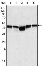
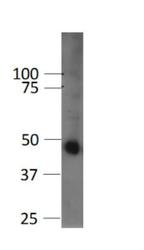
- Western Blot: GSK-3 beta Antibody (3D10) [NBP1-47470] - Analysis using GSK3 beta mouse mAb against A549 (1), K562 (2), PC-12 (3), NIH/3T3 (4), and HEK293 (5) cell lysates.

- Submitted by
- Novus Biologicals (provider)
- Main image
- Experimental details
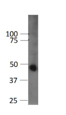
- Western Blot: GSK-3 beta Antibody (3D10) [NBP1-47470] - Analysis of GSK-3 beta in mouse beta cell line (betaTC3) using anti-GSK-3 beta antibody. Image from verified customer review.
Supportive validation
- Submitted by
- Novus Biologicals (provider)
- Main image
- Experimental details
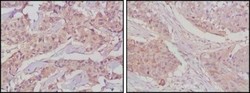
- Immunohistochemistry-Paraffin: GSK-3 beta Antibody (3D10) [NBP1-47470] - Analysis of paraffin-embedded human lung cancer (left) and breast cancer tissues (right) using GSK3 beta mouse mAb with DAB staining.

Supportive validation
- Submitted by
- Novus Biologicals (provider)
- Main image
- Experimental details
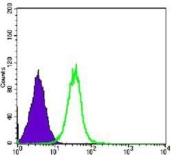
- Flow Cytometry: GSK-3 beta Antibody (3D10) [NBP1-47470] - Flow cytometric analysis of Hela cells using GSK3 beta mouse mAb (green) and negative control (purple).

- Submitted by
- Novus Biologicals (provider)
- Main image
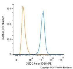
- Experimental details
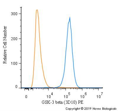
- Flow Cytometry: GSK-3 beta Antibody (3D10) [NBP1-47470] - An intracellular stain was performed on HeLa cells with GSK-3 beta (3D10) antibody NBP1-47470PE (blue) and a matched isotype control (orange). Cells were fixed with 4% PFA and then permeablized with 0.1% saponin. Cells were incubated in an antibody dilution of 2.5 ug/mL for 30 minutes at room temperature. Both antibodies were conjugated to Phycoerythrin.