Explore
 Validate
 Learn
Learn Western blot
 Immunocytochemistry
Antibody data
- Antibody Data
- Antigen structure
- References [1]
- Comments [0]
- Validations
- Immunocytochemistry [2]
- Flow cytometry [2]
- Other assay [1]
Submit
Validation data
Reference
Comment
Report error
- Product number
- MA1-206 - Provider product page

- Provider
- Invitrogen Antibodies
- Product name
- CXCR1 Monoclonal Antibody (501)
- Antibody type
- Monoclonal
- Antigen
- Synthetic peptide
- Description
- MA1-206 detects CXCR1/IL8RA from human samples. MA1-206 has been successfully tested in western blot, immunofluorescence and flow cytometry applications. In western blot, MA1-206 detects CXCR1/IL8RA (human) at approximate molecular weight of 30 kDa.
- Reactivity
- Human
- Host
- Mouse
- Isotype
- IgG
- Antibody clone number
- 501
- Vial size
- 100 μg
- Concentration
- 1 mg/mL
- Storage
- -20°C, Avoid Freeze/Thaw Cycles
Submitted references Overexpression of BQ323636.1 Modulated AR/IL-8/CXCR1 Axis to Confer Tamoxifen Resistance in ER-Positive Breast Cancer.
Tsoi H, Shi L, Leung MH, Man EPS, So ZQ, Chan WL, Khoo US
Life (Basel, Switzerland) 2022 Jan 10;12(1)
Life (Basel, Switzerland) 2022 Jan 10;12(1)
No comments: Submit comment
Supportive validation
- Submitted by
- Invitrogen Antibodies (provider)
- Main image

- Experimental details
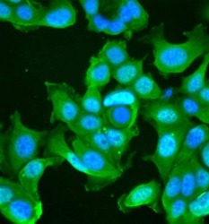
- Immunofluorescent analysis of CXCR1/IL8RA (green) in MCF7 cells. Cells were blocked with blocking buffer (Product # 37525) for 30 min at room temparture and then stained with CXCR1/IL8RA mouse monoclonal antibody (Product # MA1-206) at a dilution of 1:100 for 1 hour at room temperature. After washing with PBS for 3 times, the cells were then stained with Alexa Fluor 488 goat anti-mouse secondary antibody (Product # A28175) for 1 hour at room temperature. The cells were fixed with 4% paraformaldehyde for 30 minutes at room temperature. Nuclei (blue) were counterstained with Hoechst 33342 dye (Product # 62249). Images were taken on a Thermo Scientific EVOS FL Cell Imaging System at 40X magnification.

- Submitted by
- Invitrogen Antibodies (provider)
- Main image
- Experimental details
- Immunofluorescent analysis of CXCR1/IL8RA (green) in MCF7 cells. Cells were blocked with blocking buffer (Product # 37525) for 30 min at room temparture and then stained with CXCR1/IL8RA mouse monoclonal antibody (Product # MA1-206) at a dilution of 1:100 for 1 hour at room temperature. After washing with PBS for 3 times, the cells were then stained with Alexa Fluor 488 goat anti-mouse secondary antibody (Product # A28175) for 1 hour at room temperature. The cells were fixed with 4% paraformaldehyde for 30 minutes at room temperature. Nuclei (blue) were counterstained with Hoechst 33342 dye (Product # 62249). Images were taken on a Thermo Scientific EVOS FL Cell Imaging System at 40X magnification.

Supportive validation
- Submitted by
- Invitrogen Antibodies (provider)
- Main image
- Experimental details
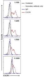
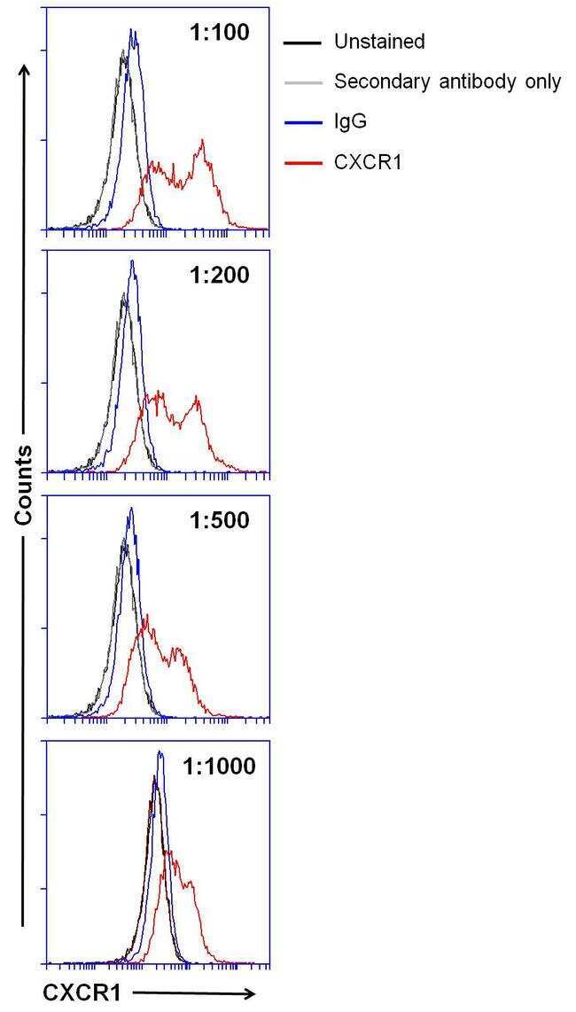
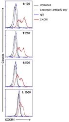
- Flow cytometry analysis of CXCR1/IL8RA on Raji cells. Cells were blocked with blocking buffer (Product # 37525) for 30 min at room temparture and then stained with CXCR1/IL8RA mouse monoclonal antibody (Product # MA1-206) at a dilution as indicated for 1 hour on ice. After washing with ice-cold PBS for 3 times, the cells were stained with Alexa Fluor 488 goat anti-mouse secondary antibody (Product # A28175) for 1 hour on ice. A representative 10,000 cells were acquired for each sample.
- Submitted by
- Invitrogen Antibodies (provider)
- Main image
- Experimental details
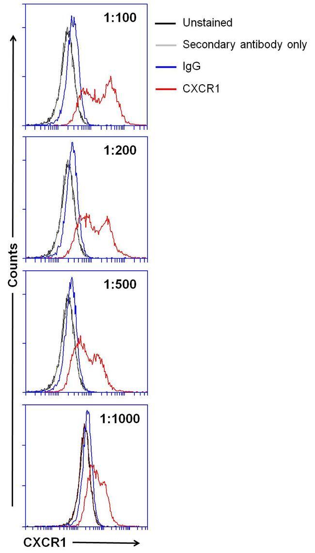
- Flow cytometry analysis of CXCR1/IL8RA on Raji cells. Cells were blocked with blocking buffer (Product # 37525) for 30 min at room temparture and then stained with CXCR1/IL8RA mouse monoclonal antibody (Product # MA1-206) at a dilution as indicated for 1 hour on ice. After washing with ice-cold PBS for 3 times, the cells were stained with Alexa Fluor 488 goat anti-mouse secondary antibody (Product # A28175) for 1 hour on ice. A representative 10,000 cells were acquired for each sample.
Supportive validation
- Submitted by
- Invitrogen Antibodies (provider)
- Main image
- Experimental details
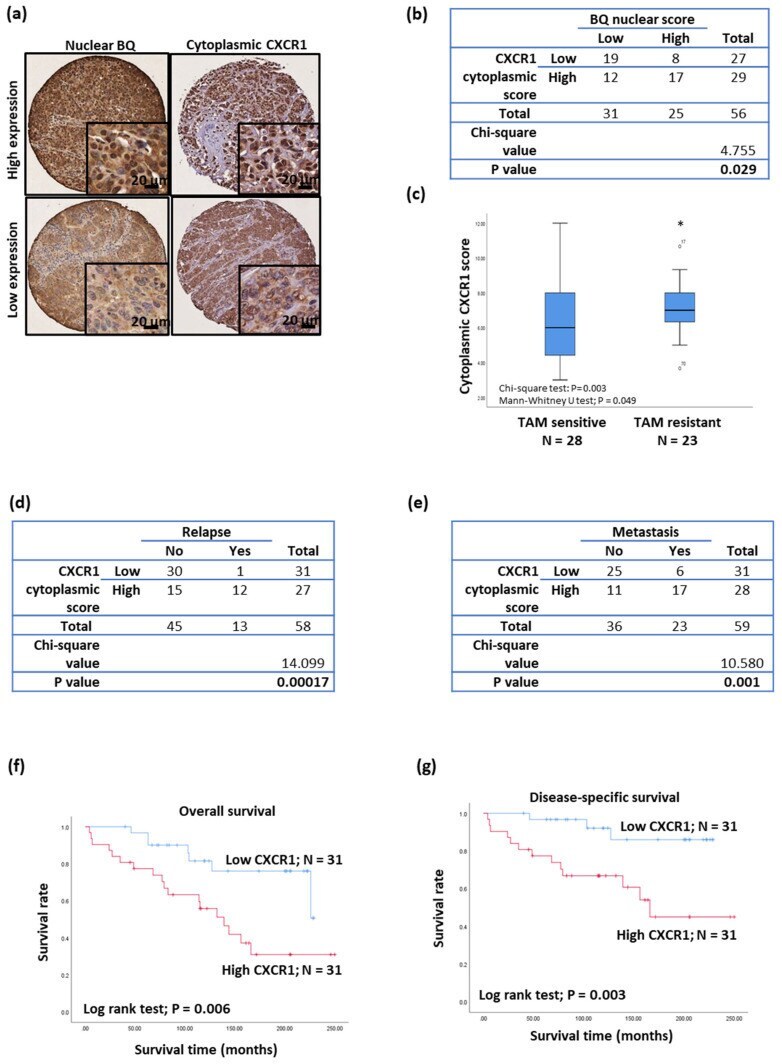
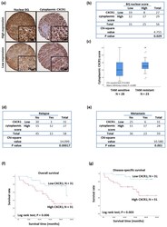
- Figure 8 Clinical significance of CXCR1 in breast cancer. ( a ) Immunohistochemistry of BQ and CXCR2 was performed on primary ER+ve breast tumor on TMA. ( b ) Chi-square test to determine the correlation between nuclear BQ and cytoplasmic CXCR1. ( c ) Tamoxifen resistance was associated with high expression of cytoplasmic CXCR1. Chi-square test and Mann-Whitney U test were employed. * represents p < 0.05. Chi-square test to determine the correlation of cytoplasmic CXCR1 with ( d ) relapse and ( e ) metastasis. High expression of CXCR1 was associated with poorer ( f ) overall survival and ( g ) disease-specific survival in ER+ve breast cancer. Log-rank test was employed.