 Explore
 Validate
 Learn
Learn Western blot
 Immunocytochemistry
Antibody data
- Antibody Data
- Antigen structure
- References [0]
- Comments [0]
- Validations
- Immunocytochemistry [4]
- Immunoprecipitation [1]
- Flow cytometry [2]
Submit
Validation data
Reference
Comment
Report error
- Product number
- MA5-14893 - Provider product page

- Provider
- Invitrogen Antibodies
- Product name
- SHIP1 Monoclonal Antibody (T.7.7)
- Antibody type
- Monoclonal
- Antigen
- Synthetic peptide
- Description
- It is not recommended to aliquot this antibody.
- Reactivity
- Human
- Host
- Rabbit
- Isotype
- IgG
- Antibody clone number
- T.7.7
- Vial size
- 100 μL
- Concentration
- 44 μg/mL
- Storage
- -20°C
No comments: Submit comment
Supportive validation
- Submitted by
- Invitrogen Antibodies (provider)
- Main image

- Experimental details
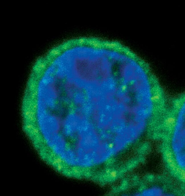
- Immunofluorescent analysis of SHIP1 in RL cells using a SHIP1 monoclonal antibody (Product # MA5-14893) (green). DNA is labeled using a fluorescent blue dye.
- Submitted by
- Invitrogen Antibodies (provider)
- Main image

- Experimental details
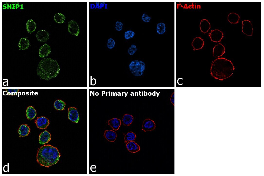
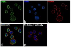
- Immunofluorescence analysis of SHIP1 was performed using 70% confluent log phase THP-1 cells. The cells were fixed with 4% paraformaldehyde for 10 minutes, permeabilized with 0.1% Triton™ X-100 for 15 minutes, and blocked with 2% BSA for 1 hour at room temperature. The cells were labeled with SHIP1 Rabbit Monoclonal Antibody (T.7.7) (Product # MA5-14893) at 1:100 dilution in 0.1% BSA, incubated at 4 degree Celsius overnight and then with Goat anti-Rabbit IgG (H+L) Highly Cross-Adsorbed Secondary Antibody, Alexa Fluor Plus 488 (Product # A32731) at a dilution of 1:2000 for 45 minutes at room temperature (Panel a: green). Nuclei (Panel b: blue) were stained with ProLong™ Diamond Antifade Mountant with DAPI (Product # P36962). F-actin (Panel c: red) was stained with Rhodamine Phalloidin (Product # R415, 1:300). Panel d represents the merged image showing cytoplasmic localization. Panel e represents control cells with no primary antibody to assess background. The images were captured at 60X magnification.
- Submitted by
- Invitrogen Antibodies (provider)
- Main image

- Experimental details
- Immunofluorescent analysis of SHIP1 in RL cells using a SHIP1 monoclonal antibody (Product # MA5-14893) (green). DNA is labeled using a fluorescent blue dye.

- Submitted by
- Invitrogen Antibodies (provider)
- Main image
- Experimental details
- Immunofluorescence analysis of SHIP1 was performed using 70% confluent log phase THP-1 cells. The cells were fixed with 4% paraformaldehyde for 10 minutes, permeabilized with 0.1% Triton™ X-100 for 15 minutes, and blocked with 2% BSA for 1 hour at room temperature. The cells were labeled with SHIP1 Rabbit Monoclonal Antibody (T.7.7) (Product # MA5-14893) at 1:100 dilution in 0.1% BSA, incubated at 4 degree Celsius overnight and then with Goat anti-Rabbit IgG (H+L) Highly Cross-Adsorbed Secondary Antibody, Alexa Fluor Plus 488 (Product # A32731) at a dilution of 1:2000 for 45 minutes at room temperature (Panel a: green). Nuclei (Panel b: blue) were stained with ProLong™ Diamond Antifade Mountant with DAPI (Product # P36962). F-actin (Panel c: red) was stained with Rhodamine Phalloidin (Product # R415, 1:300). Panel d represents the merged image showing cytoplasmic localization. Panel e represents control cells with no primary antibody to assess background. The images were captured at 60X magnification.

Supportive validation
- Submitted by
- Invitrogen Antibodies (provider)
- Main image
- Experimental details
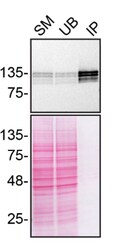
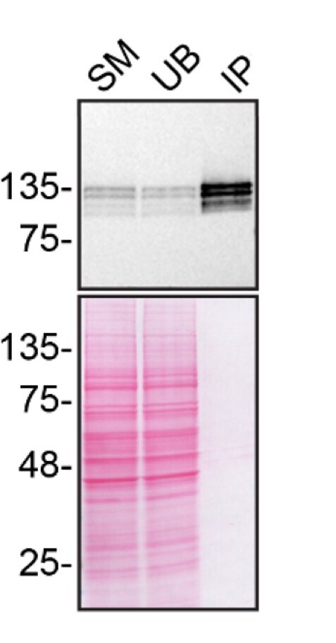
- Immunoprecipitation of SHIP1 was performed on U-937 WT cell lysate. Antibody-bead conjugate was prepared by adding 2 µg of SHIP1 Monoclonal Antibody (T.7.7) (Product # MA5-14893) and 30 µL of Dynabeads™ Protein A (Product # 10002D) to 500 µl of Pierce™ IP Lysis Buffer (Product # 87788). The mixture was rocked for ~1 hour at 4 degree celcius followed by two washes to remove unbound antibodies. U-937 WT cells were lysed in Pierce™ IP Lysis Buffer (Product # 87788) supplemented with protease inhibitor. The lysate was rocked for 30 min at 4 degree celcius and spun at 110,000xg for 15 min at 4 degree celcius. One mg of lysate was incubated with the antibody-bead conjugate for ~1 hours at 4 degree celcius. Following centrifugation and multiple washes, 4% starting material (SM), 4% unbound fraction (UB) and immunoprecipitated fraction (IP) were processed for immunoblot using a different antibody. Ponceau stained transfer of blot is shown (below immunoblot). Data courtesy of YCharOS Inc., an open science company with the mission of characterizing commercially available antibodies using knockout validation.
Supportive validation
- Submitted by
- Invitrogen Antibodies (provider)
- Main image
- Experimental details
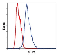
- Flow cytometric analysis of SHIP1 in Jurkat cells (red) and RL cells (blue) using a SHIP1 monoclonal antibody (Product # MA5-14893).

- Submitted by
- Invitrogen Antibodies (provider)
- Main image
- Experimental details
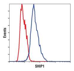
- Flow cytometric analysis of SHIP1 in Jurkat cells (red) and RL cells (blue) using a SHIP1 monoclonal antibody (Product # MA5-14893).
