Explore
 Validate
 Learn
Learn Western blot
 Immunocytochemistry
Antibody data
- Antibody Data
- Antigen structure
- References [3]
- Comments [0]
- Validations
- Immunocytochemistry [4]
- Other assay [1]
Submit
Validation data
Reference
Comment
Report error
- Product number
- MA1-177 - Provider product page

- Provider
- Invitrogen Antibodies
- Product name
- BACE1 Monoclonal Antibody (3C1C3)
- Antibody type
- Monoclonal
- Antigen
- Purifed from natural sources
- Description
- MA1-177 targets BACE1 in WB and IF, shows reactivity with Human, Primate, Canine, Mouse and Rat samples.
- Reactivity
- Human, Mouse, Rat, Canine
- Host
- Mouse
- Isotype
- IgG
- Antibody clone number
- 3C1C3
- Vial size
- 100 μg
- Concentration
- 1 mg/mL
- Storage
- -20°C, Avoid Freeze/Thaw Cycles
Submitted references Acidifying Endolysosomes Prevented Low-Density Lipoprotein-Induced Amyloidogenesis.
Chenodeoxycholic Acid Ameliorates AlCl(3)-Induced Alzheimer's Disease Neurotoxicity and Cognitive Deterioration via Enhanced Insulin Signaling in Rats.
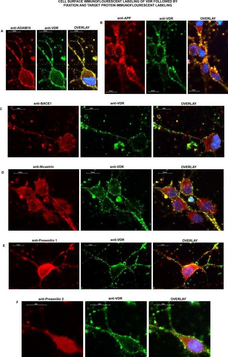
Vitamin D receptor is present on the neuronal plasma membrane and is co-localized with amyloid precursor protein, ADAM10 or Nicastrin.
Hui L, Soliman ML, Geiger NH, Miller NM, Afghah Z, Lakpa KL, Chen X, Geiger JD
Journal of Alzheimer's disease : JAD 2019;67(1):393-410
Journal of Alzheimer's disease : JAD 2019;67(1):393-410
Chenodeoxycholic Acid Ameliorates AlCl(3)-Induced Alzheimer's Disease Neurotoxicity and Cognitive Deterioration via Enhanced Insulin Signaling in Rats.
Bazzari FH, Abdallah DM, El-Abhar HS
Molecules (Basel, Switzerland) 2019 May 24;24(10)
Molecules (Basel, Switzerland) 2019 May 24;24(10)
Vitamin D receptor is present on the neuronal plasma membrane and is co-localized with amyloid precursor protein, ADAM10 or Nicastrin.
Dursun E, Gezen-Ak D
PloS one 2017;12(11):e0188605
PloS one 2017;12(11):e0188605
No comments: Submit comment
Supportive validation
- Submitted by
- Invitrogen Antibodies (provider)
- Main image
- Experimental details
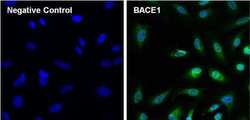
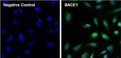
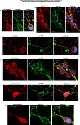
- Immunofluorescent analysis of BACE1 (green) in HeLa cells. The cells were fixed with formalin for 15 minutes, permeabilized with 0.1% Triton X-100 in TBS for 10 minutes, and blocked with 5% Normal Goat Serum (Product # 31872) for 15 minutes at room temperature. Cells were stained with or without BACE1 monoclonal antibody (Product # MA1-177), at a dilution of 1:50 overnight at 4C, and then incubated with a DyLight 488 goat anti-mouse IgG secondary antibody (Product # 35503) at a dilution of 1:1000 for 30 minutes at room temperature (both panels, green). Nuclei (both panels, blue) were stained with Hoechst 33342 dye (Product # 62249). Images were taken on a Thermo Scientific ToxInsight at 20X magnification.

- Submitted by
- Invitrogen Antibodies (provider)
- Main image

- Experimental details
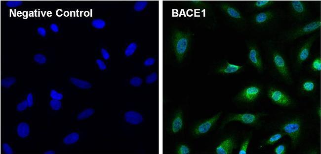
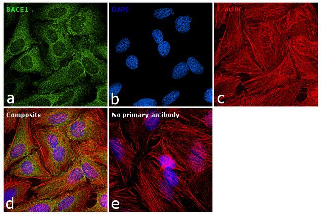
- Immunofluorescence analysis of BACE1 was performed using 70% confluent log phase HeLa cells. The cells were fixed with 4% paraformaldehyde for 10 minutes, permeabilized with 0.1% Triton™ X-100 for 15 minutes, and blocked with 1% BSA for 1 hour at room temperature. The cells were labeled with BACE1 Monoclonal Antibody (3C1C3) (Product # MA1-177) at 1:100 dilution in 0.1% BSA, incubated at 4 degree Celsius overnight and then labeled with Goat anti-Mouse IgG (H+L) Superclonal™ Secondary Antibody, Alexa Fluor® 488 conjugate (Product # A28175) at a dilution of 1:2000 for 45 minutes at room temperature (Panel a: green). Nuclei (Panel b: blue) were stained with ProLong™ Diamond Antifade Mountant with DAPI (Product # P36962). F-actin (Panel c: red) was stained with Rhodamine Phalloidin (Product # R415). Panel d represents the merged image showing cytoplasmic localization. The images were captured at 60X magnification.

- Submitted by
- Invitrogen Antibodies (provider)
- Main image
- Experimental details
- Immunofluorescent analysis of BACE1 (green) in HeLa cells. The cells were fixed with formalin for 15 minutes, permeabilized with 0.1% Triton X-100 in TBS for 10 minutes, and blocked with 5% Normal Goat Serum (Product # 31872) for 15 minutes at room temperature. Cells were stained with or without BACE1 monoclonal antibody (Product # MA1-177), at a dilution of 1:50 overnight at 4C, and then incubated with a DyLight 488 goat anti-mouse IgG secondary antibody (Product # 35503) at a dilution of 1:1000 for 30 minutes at room temperature (both panels, green). Nuclei (both panels, blue) were stained with Hoechst 33342 dye (Product # 62249). Images were taken on a Thermo Scientific ToxInsight at 20X magnification.
- Submitted by
- Invitrogen Antibodies (provider)
- Main image

- Experimental details
- Immunofluorescence analysis of BACE1 was performed using 70% confluent log phase HeLa cells. The cells were fixed with 4% paraformaldehyde for 10 minutes, permeabilized with 0.1% Triton™ X-100 for 15 minutes, and blocked with 1% BSA for 1 hour at room temperature. The cells were labeled with BACE1 Monoclonal Antibody (3C1C3) (Product # MA1-177) at 1:100 dilution in 0.1% BSA, incubated at 4 degree Celsius overnight and then labeled with Goat anti-Mouse IgG (H+L) Superclonal™ Secondary Antibody, Alexa Fluor® 488 conjugate (Product # A28175) at a dilution of 1:2000 for 45 minutes at room temperature (Panel a: green). Nuclei (Panel b: blue) were stained with ProLong™ Diamond Antifade Mountant with DAPI (Product # P36962). F-actin (Panel c: red) was stained with Rhodamine Phalloidin (Product # R415). Panel d represents the merged image showing cytoplasmic localization. The images were captured at 60X magnification.
Supportive validation
- Submitted by
- Invitrogen Antibodies (provider)
- Main image
- Experimental details
- NULL