 Explore
 Validate
 Learn
Learn Western blot
Antibody data
- Antibody Data
- Antigen structure
- References [0]
- Comments [0]
- Validations
- Western blot [2]
- Immunocytochemistry [5]
- Immunohistochemistry [1]
Submit
Validation data
Reference
Comment
Report error
- Product number
- PA5-19952 - Provider product page

- Provider
- Invitrogen Antibodies
- Product name
- BACE1 Polyclonal Antibody
- Antibody type
- Polyclonal
- Antigen
- Synthetic peptide
- Description
- A suggested positive control is human brain tissue lysate. PA5-19952 can be used with blocking peptide PEP-0077. The PA5-19952 immunogen is located within the last 50 amino acids of BACE. Predicted molecular ~ 55kD. In Western blot applications, this antibody has been observed to detect a band at: 65kD (Post-modification: 4 N-linked glycosylation). Predicted species reactivity based on immunogen sequence: Guinea pig: (94%), Rat: (94%), Bovine: (94%).
- Reactivity
- Human, Mouse
- Host
- Rabbit
- Isotype
- IgG
- Vial size
- 100 μg
- Concentration
- 1 mg/mL
- Storage
- Maintain refrigerated at 2-8°C for up to 3 months. For long term storage store at -20°C
No comments: Submit comment
Supportive validation
- Submitted by
- Invitrogen Antibodies (provider)
- Main image

- Experimental details
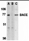
- Western Blot Validation of BACE. Loading: 15 µg of lysates per lane. Antibodies: BACE1 Polyclonal Antibody (Product # PA5-19952) (1 µg/mL), 1h incubation at RT in 0.05 NFDM/TBST. Secondary: Goat anti-rabbit IgG HRP conjugate at 1:10,000 dilution. Lane A-C: human brain tissue lysate in the absence (A) or presence (B) of blocking peptide and mouse 3T3/NIH cell lysate (C).
- Submitted by
- Invitrogen Antibodies (provider)
- Main image

- Experimental details
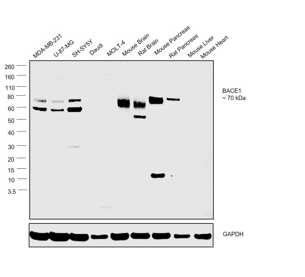
- Western blot was performed using Anti-BACE1 Polyclonal Antibody(Product # PA5-19952) and a 70 kDa band corresponding to BACE1 was observed across all the cell lines and tissues tested except Daudi, MOLT-4, Mouse Liver and Mouse Heart. Whole cell extracts (30 µg lysate) of MDA-MB-231 (Lane 1), U-87-MG (Lane 2), SH-SY-5Y (Lane 3), Daudi (Lane 4), MOLT-4 (Lane 5), Mouse Brain (Lane 6), Rat Brain (Lane 7), Mouse Pancreas (Lane 8), Rat Pancreas (Lane 9), Mouse Liver (Lane 10) and Mouse Heart (Lane 11) were electrophoresed using NuPAGE™ 4-12% Bis-Tris Protein Gel (Product # NP0322BOX). Resolved proteins were then transferred onto a nitrocellulose membrane (Product # IB23001) by iBlot® 2 Dry Blotting System (Product # IB21001). The blot was probed with the primary antibody (1 µg/mL) and detected by chemiluminescence with Goat anti-Rabbit IgG (Heavy Chain), Superclonal™ Recombinant Secondary Antibody, HRP (Product # A27036, 1:4,000 dilution) using the iBright FL 1000 (Product # A32752). Chemiluminescent detection was performed using Novex® ECL Chemiluminescent Substrate Reagent Kit (Product # WP20005).
Supportive validation
- Submitted by
- Invitrogen Antibodies (provider)
- Main image

- Experimental details
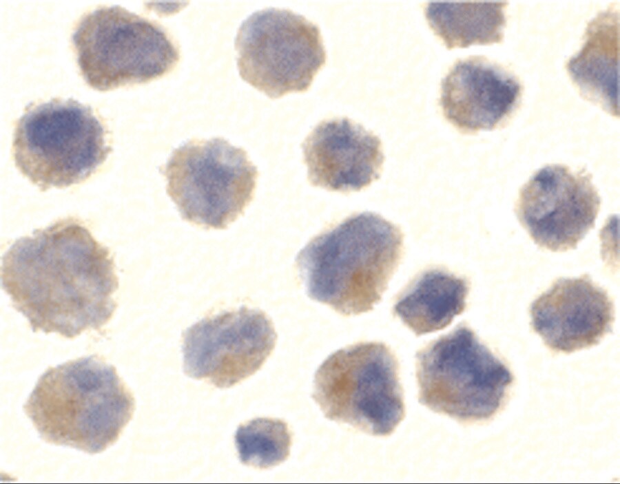
- Immunocytochemistry of 3T3/NIH cells using BACE1 Polyclonal Antibody (Product # PA5-19952) at 10 µg/mL. Cells were fixed with formaldehyde and blocked with 0.1 serum for 1 h at RT; antigen retrieval was by heat mediation with a citrate buffer (pH6). Samples were incubated with primary antibody overnight at 4°C. A goat anti-rabbit IgG H&L (HRP) at 1:250 was used as secondary. Counter stained with Hematoxylin.
- Submitted by
- Invitrogen Antibodies (provider)
- Main image

- Experimental details
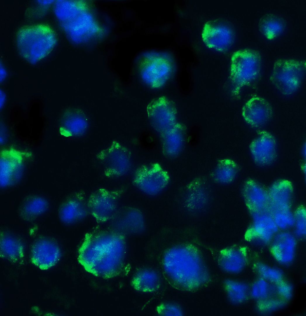
- Immunofluorescent analysis of 4% paraformaldehyde-fixed mouse 3T3/NIH cells labeling BACE with BACE1 Polyclonal Antibody (Product # PA5-19952) at 20 µg/mL, followed by goat anti-rabbit IgG secondary antibody at 1:500 dilution (green) and DAPI staining (blue). Image showing both membrane and cytosol staining on 3T3/NIH cells.
- Submitted by
- Invitrogen Antibodies (provider)
- Main image

- Experimental details
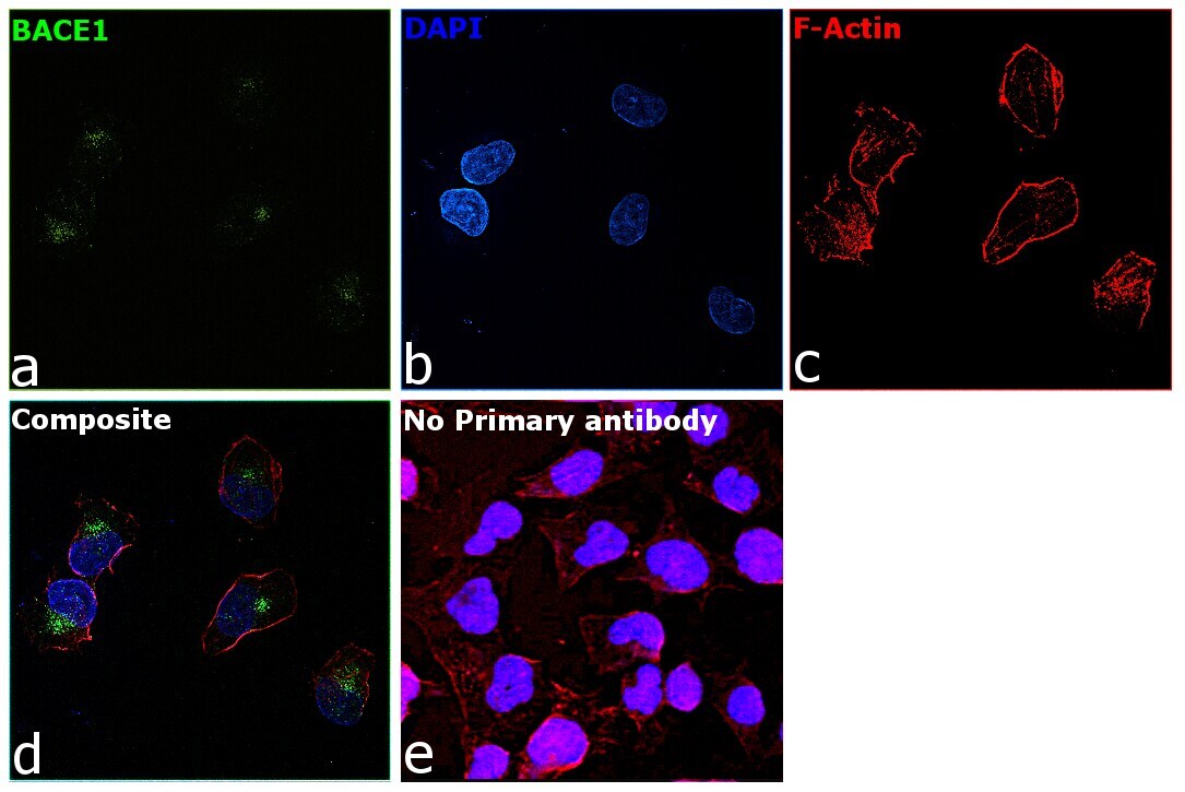
- Immunofluorescence analysis of BACE1 was performed using 70% confluent log phase MDA-MB-231 cells. The cells were fixed with 4% Paraformaldehyde for 10 Minutes, permeabilized with 0.1% Triton™ X-100 for 10 Minutes, and blocked with 2% BSA for 10 Minutes at room temperature. The cells were labeled with BACE1 Polyclonal Antibody (Product # PA5-19952) at 10 µg/mL in 0.1% BSA, incubated at 4 degree Celsius overnight and then labeled with Goat anti-Rabbit IgG (H+L) Superclonal™ Recombinant Secondary Antibody, Alexa Fluor® 488 conjugate (Product # A27034, 1:2,000 dilution) for 45 minutes at room temperature (Panel a: Green). Nuclei (Panel b: Blue) were stained with SlowFade® Gold Antifade Mountant with DAPI (Product # S36938). F-actin (Panel c: Red) was stained with Rhodamine Phalloidin (Product # R415, 1:300). Panel d represents the merged image showing golgi like cytoplasmic localization. Panel e represents control cells with no primary antibody to assess background. The images were captured at 60X magnification.
- Submitted by
- Invitrogen Antibodies (provider)
- Main image

- Experimental details
- Immunofluorescent analysis of 4% paraformaldehyde-fixed mouse 3T3/NIH cells labeling BACE with BACE1 Polyclonal Antibody (Product # PA5-19952) at 20 µg/mL, followed by goat anti-rabbit IgG secondary antibody at 1:500 dilution (green) and DAPI staining (blue). Image showing both membrane and cytosol staining on 3T3/NIH cells.

- Submitted by
- Invitrogen Antibodies (provider)
- Main image

- Experimental details
- Immunofluorescence analysis of BACE1 was performed using 70% confluent log phase MDA-MB-231 cells. The cells were fixed with 4% Paraformaldehyde for 10 Minutes, permeabilized with 0.1% Triton™ X-100 for 10 Minutes, and blocked with 2% BSA for 10 Minutes at room temperature. The cells were labeled with BACE1 Polyclonal Antibody (Product # PA5-19952) at 10 µg/mL in 0.1% BSA, incubated at 4 degree Celsius overnight and then labeled with Goat anti-Rabbit IgG (Heavy Chain) Superclonal™ Recombinant Secondary Antibody, Alexa Fluor® 488 conjugate (Product # A27034, 1:2,000 dilution) for 45 minutes at room temperature (Panel a: Green). Nuclei (Panel b: Blue) were stained with SlowFade® Gold Antifade Mountant with DAPI (Product # S36938). F-actin (Panel c: Red) was stained with Rhodamine Phalloidin (Product # R415, 1:300). Panel d represents the merged image showing golgi like cytoplasmic localization. Panel e represents control cells with no primary antibody to assess background. The images were captured at 60X magnification.

Supportive validation
- Submitted by
- Invitrogen Antibodies (provider)
- Main image

- Experimental details
- Immunohistochemical analysis of paraffin-embedded mouse brain tissue using BACE1 Polyclonal Antibody (Product # PA5-19952) at 2.5 µg/mL. Tissue was fixed with formaldehyde and blocked with 0.1 serum for 1 h at RT; antigen retrieval was by heat mediation with a citrate buffer (pH6). Samples were incubated with primary antibody overnight at 4˚C. A goat anti-rabbit IgG H&L (HRP) at 1/250 was used as secondary. Counter stained with Hematoxylin.
