Explore
 Validate
 Learn
Learn Western blot
 Immunohistochemistry
Antibody data
- Antibody Data
- Antigen structure
- References [16]
- Comments [0]
- Validations
- Immunohistochemistry [1]
- Other assay [9]
Submit
Validation data
Reference
Comment
Report error
- Product number
- PA1-1746 - Provider product page

- Provider
- Invitrogen Antibodies
- Product name
- Aggrecan Neo Polyclonal Antibody
- Antibody type
- Polyclonal
- Antigen
- Synthetic peptide
- Description
- PA1-1746 detects human, rat, mouse, bovine, canine, and porcine aggrecan neo G1. PA1-1746 has been successfully used in Western blot and immunofluorescence procedures. By Western blot, this antibody detects an ~64 kDa protein representing the neoepitope sequence (NITEGE) generated by aggrecanase-mediated cleavage at Glu373-Ala374 of aggrecan core proteins. The PA1-1746 immunogen is a synthetic peptide corresponding to residues residues C G G N(387) I T E G E(392) of human aggrecan.
- Reactivity
- Human, Mouse, Rat, Bovine, Canine, Porcine
- Host
- Rabbit
- Isotype
- IgG
- Vial size
- 100 μg
- Concentration
- 0.83 mg/mL
- Storage
- -20°C, Avoid Freeze/Thaw Cycles
Submitted references Role of Ciliary Protein Intraflagellar Transport Protein 88 in the Regulation of Cartilage Thickness and Osteoarthritis Development in Mice.
Platelet-Derived Growth Factor-BB Inhibits Intervertebral Disc Degeneration via Suppressing Pyroptosis and Activating the MAPK Signaling Pathway.
Secreted frizzled-related protein 3 was genetically and functionally associated with developmental dysplasia of the hip.
Vascular dimorphism ensured by regulated proteoglycan dynamics favors rapid umbilical artery closure at birth.
Glycoproteomic Analysis of the Aortic Extracellular Matrix in Marfan Patients.
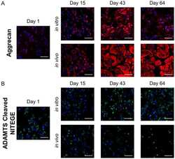
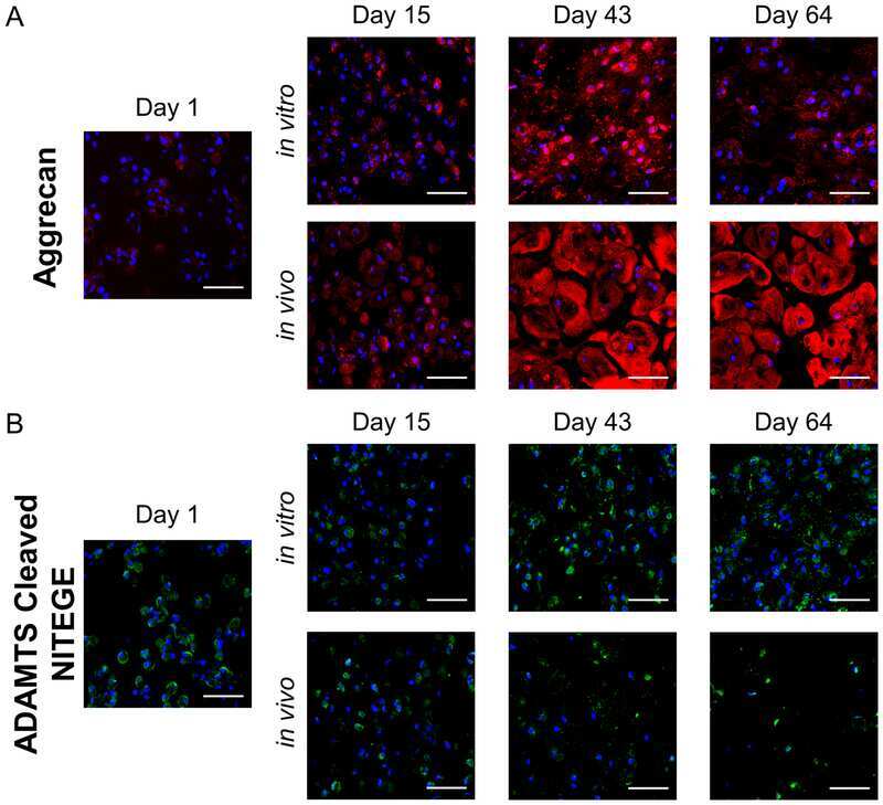
An in vitro and in vivo comparison of cartilage growth in chondrocyte-laden matrix metalloproteinase-sensitive poly(ethylene glycol) hydrogels with localized transforming growth factor β3.
Extracellular Matrix Proteomics Reveals Interplay of Aggrecan and Aggrecanases in Vascular Remodeling of Stented Coronary Arteries.
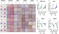
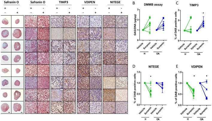
Suramin increases cartilage proteoglycan accumulation in vitro and protects against joint damage triggered by papain injection in mouse knees in vivo.
Olive and grape seed extract prevents post-traumatic osteoarthritis damages and exhibits in vitro anti IL-1β activities before and after oral consumption.
Brief Report: JNK-2 Controls Aggrecan Degradation in Murine Articular Cartilage and the Development of Experimental Osteoarthritis.
Transient anabolic effects accompany epidermal growth factor receptor signal activation in articular cartilage in vivo.
Adamts5 (aggrecanase-2) is widely expressed in the mouse musculoskeletal system and is induced in specific regions of knee joint explants by inflammatory cytokines.
Transcription factor Nfat1 deficiency causes osteoarthritis through dysfunction of adult articular chondrocytes.
Analysis of aggrecan in human knee cartilage and synovial fluid indicates that aggrecanase (ADAMTS) activity is responsible for the catabolic turnover and loss of whole aggrecan whereas other protease activity is required for C-terminal processing in vivo.
The intermediates of aggrecanase-dependent cleavage of aggrecan in rat chondrosarcoma cells treated with interleukin-1.
The intermediates of aggrecanase-dependent cleavage of aggrecan in rat chondrosarcoma cells treated with interleukin-1.
Coveney CR, Zhu L, Miotla-Zarebska J, Stott B, Parisi I, Batchelor V, Duarte C, Chang E, McSorley E, Vincent TL, Wann AKT
Arthritis & rheumatology (Hoboken, N.J.) 2022 Jan;74(1):49-59
Arthritis & rheumatology (Hoboken, N.J.) 2022 Jan;74(1):49-59
Platelet-Derived Growth Factor-BB Inhibits Intervertebral Disc Degeneration via Suppressing Pyroptosis and Activating the MAPK Signaling Pathway.
Zhang W, Gong Y, Zheng X, Qiu J, Jiang T, Chen L, Lu F, Wu X, Cheng F, Hong Z
Frontiers in pharmacology 2021;12:799130
Frontiers in pharmacology 2021;12:799130
Secreted frizzled-related protein 3 was genetically and functionally associated with developmental dysplasia of the hip.
Xu R, Zhang F, Lu J, Wang K, Pan P, Sun Y, Zhang Y
Aging 2021 Apr 4;13(8):11281-11295
Aging 2021 Apr 4;13(8):11281-11295
Vascular dimorphism ensured by regulated proteoglycan dynamics favors rapid umbilical artery closure at birth.
Nandadasa S, Szafron JM, Pathak V, Murtada SI, Kraft CM, O'Donnell A, Norvik C, Hughes C, Caterson B, Domowicz MS, Schwartz NB, Tran-Lundmark K, Veigl M, Sedwick D, Philipson EH, Humphrey JD, Apte SS
eLife 2020 Sep 10;9
eLife 2020 Sep 10;9
Glycoproteomic Analysis of the Aortic Extracellular Matrix in Marfan Patients.
Yin 殷晓科 X, Wanga S, Fellows AL, Barallobre-Barreiro J, Lu R, Davaapil H, Franken R, Fava M, Baig F, Skroblin P, Xing Q, Koolbergen DR, Groenink M, Zwinderman AH, Balm R, de Vries CJM, Mulder BJM, Viner R, Jahangiri M, Reinhardt DP, Sinha S, de Waard V, Mayr M
Arteriosclerosis, thrombosis, and vascular biology 2019 Sep;39(9):1859-1873
Arteriosclerosis, thrombosis, and vascular biology 2019 Sep;39(9):1859-1873
An in vitro and in vivo comparison of cartilage growth in chondrocyte-laden matrix metalloproteinase-sensitive poly(ethylene glycol) hydrogels with localized transforming growth factor β3.
Schneider MC, Chu S, Randolph MA, Bryant SJ
Acta biomaterialia 2019 Jul 15;93:97-110
Acta biomaterialia 2019 Jul 15;93:97-110
Extracellular Matrix Proteomics Reveals Interplay of Aggrecan and Aggrecanases in Vascular Remodeling of Stented Coronary Arteries.
Suna G, Wojakowski W, Lynch M, Barallobre-Barreiro J, Yin X, Mayr U, Baig F, Lu R, Fava M, Hayward R, Molenaar C, White SJ, Roleder T, Milewski KP, Gasior P, Buszman PP, Buszman P, Jahangiri M, Shanahan CM, Hill J, Mayr M
Circulation 2018 Jan 9;137(2):166-183
Circulation 2018 Jan 9;137(2):166-183
Suramin increases cartilage proteoglycan accumulation in vitro and protects against joint damage triggered by papain injection in mouse knees in vivo.
Guns LA, Monteagudo S, Kvasnytsia M, Kerckhofs G, Vandooren J, Opdenakker G, Lories RJ, Cailotto F
RMD open 2017;3(2):e000604
RMD open 2017;3(2):e000604
Olive and grape seed extract prevents post-traumatic osteoarthritis damages and exhibits in vitro anti IL-1β activities before and after oral consumption.
Mével E, Merceron C, Vinatier C, Krisa S, Richard T, Masson M, Lesoeur J, Hivernaud V, Gauthier O, Abadie J, Nourissat G, Houard X, Wittrant Y, Urban N, Beck L, Guicheux J
Scientific reports 2016 Sep 19;6:33527
Scientific reports 2016 Sep 19;6:33527
Brief Report: JNK-2 Controls Aggrecan Degradation in Murine Articular Cartilage and the Development of Experimental Osteoarthritis.
Ismail HM, Miotla-Zarebska J, Troeberg L, Tang X, Stott B, Yamamoto K, Nagase H, Fosang AJ, Vincent TL, Saklatvala J
Arthritis & rheumatology (Hoboken, N.J.) 2016 May;68(5):1165-71
Arthritis & rheumatology (Hoboken, N.J.) 2016 May;68(5):1165-71
Transient anabolic effects accompany epidermal growth factor receptor signal activation in articular cartilage in vivo.
Shepard JB, Jeong JW, Maihle NJ, O'Brien S, Dealy CN
Arthritis research & therapy 2013;15(3):R60
Arthritis research & therapy 2013;15(3):R60
Adamts5 (aggrecanase-2) is widely expressed in the mouse musculoskeletal system and is induced in specific regions of knee joint explants by inflammatory cytokines.
Wylie JD, Ho JC, Singh S, McCulloch DR, Apte SS
Journal of orthopaedic research : official publication of the Orthopaedic Research Society 2012 Feb;30(2):226-33
Journal of orthopaedic research : official publication of the Orthopaedic Research Society 2012 Feb;30(2):226-33
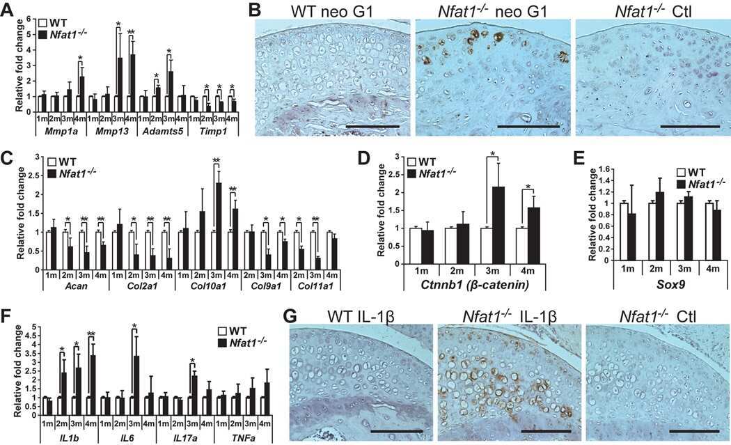
Transcription factor Nfat1 deficiency causes osteoarthritis through dysfunction of adult articular chondrocytes.
Wang J, Gardner BM, Lu Q, Rodova M, Woodbury BG, Yost JG, Roby KF, Pinson DM, Tawfik O, Anderson HC
The Journal of pathology 2009 Oct;219(2):163-72
The Journal of pathology 2009 Oct;219(2):163-72
Analysis of aggrecan in human knee cartilage and synovial fluid indicates that aggrecanase (ADAMTS) activity is responsible for the catabolic turnover and loss of whole aggrecan whereas other protease activity is required for C-terminal processing in vivo.
Sandy JD, Verscharen C
The Biochemical journal 2001 Sep 15;358(Pt 3):615-26
The Biochemical journal 2001 Sep 15;358(Pt 3):615-26
The intermediates of aggrecanase-dependent cleavage of aggrecan in rat chondrosarcoma cells treated with interleukin-1.
Sandy JD, Thompson V, Doege K, Verscharen C
The Biochemical journal 2000 Oct 1;351(Pt 1):161-6
The Biochemical journal 2000 Oct 1;351(Pt 1):161-6
The intermediates of aggrecanase-dependent cleavage of aggrecan in rat chondrosarcoma cells treated with interleukin-1.
Sandy JD, Thompson V, Doege K, Verscharen C
The Biochemical journal 2000 Oct 1;351(Pt 1):161-6
The Biochemical journal 2000 Oct 1;351(Pt 1):161-6
No comments: Submit comment
Supportive validation
- Submitted by
- Invitrogen Antibodies (provider)
- Main image
- Experimental details
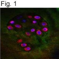
- Immunofluorescent analysis of Aggrecan Neo in OA cartilage using a polyclonal antibody (Product # PA1-1746).

Supportive validation
- Submitted by
- Invitrogen Antibodies (provider)
- Main image
- Experimental details
- NULL
- Submitted by
- Invitrogen Antibodies (provider)
- Main image
- Experimental details
- NULL
- Submitted by
- Invitrogen Antibodies (provider)
- Main image
- Experimental details
- NULL
- Submitted by
- Invitrogen Antibodies (provider)
- Main image
- Experimental details
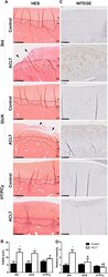
- Figure 5 HT/PCy supplementation reduces the severity of mid-stage OA in ACLT rabbits. Rabbits (n = 6 per group) underwent ACLT of the right knee. NaCl (Std), GlcN or HT/PCy were orally administrated every two days for 13 weeks starting 3 weeks before surgery. HES staining of cartilage ( A ) and OARSI scores ( B ) of non-operated knee (control) or operated knee (ACLT) at 10 weeks post-surgery are shown. Immunohistochemical detection of aggrecan epitopes (NITEGE) ( C ) and relative staining intensity of the articular cartilage matrix ( D ) of standard diet (Std), GlcN and HT/PCy diet groups at 10 weeks after ACLT. (i) Non calcified-cartilage, (ii) calcified cartilage and (iii) sub-chondral bone. *p > 0.05 compared to non-operated knee (control) within the same group; # p > 0.05 compared to ACLT receiving NaCl (Std) group, $ p > 0.05 compared to ACLT receiving GlcN. Bar represents 250 mum ( A ) and 100 mum ( C ).
- Submitted by
- Invitrogen Antibodies (provider)
- Main image

- Experimental details
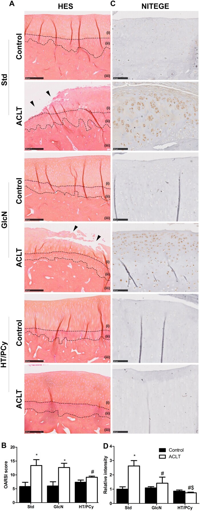
- Figure 2. ADAMTS proteoglycanases are highly expressed and active in the human umbilical vein. ( a ) RNA in situ hybridization shows robust ADAMTS1 and ADAMTS9 expression in umbilical vein endothelium and tunica media (TM) and in outer arterial TM. Robust ADAMTS4 and ADAMTS5 expression was confined to the venous endothelium, with moderate ADAMTS4 expression and minimal ADAMTS5 expression in SMC (n = 3 umbilical cords for each probe). ( b ) ADAMTS-cleaved aggrecan (anti-NITEGE, red) and versican (anti-DPEAAE, red) both showed strong ADAMTS proteolytic activity throughout the venous wall and the outer artery TM. Unlike aggrecan, extensive versican proteolysis is seen in the arterial intima and sub-intima (n = 4 umbilical cords for each antibody). Wj, Wharton''s jelly. The brackets mark TM boundaries. Scale bars in a-b = 100 mum.
- Submitted by
- Invitrogen Antibodies (provider)
- Main image
- Experimental details
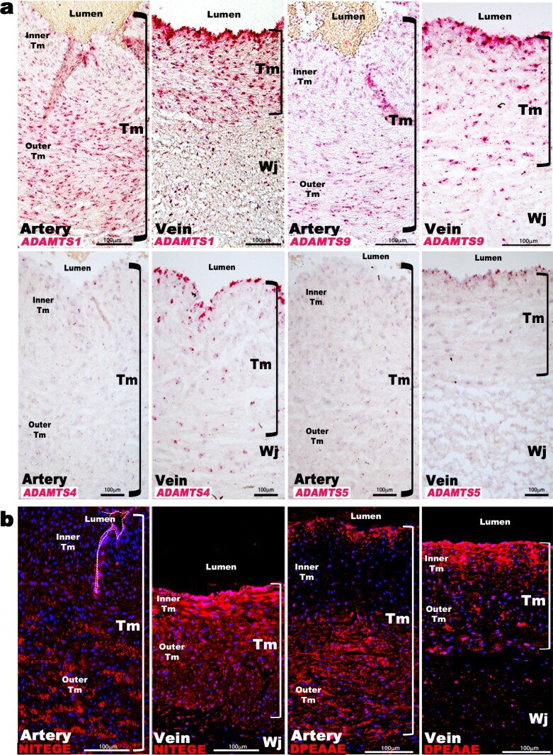
- Figure 3. Aggrecan enrichment in the inner umbilical artery tunica media (TM) and its proteolysis in the umbilical vein is a characteristic of large mammals. ( a ) Alcian blue-eosin staining of umbilical cord sections shows proteoglycan enrichment (blue) in the inner arterial tunica media (TM). The elephant umbilical vein was unavailable. ( b ) Anti-CS immunofluorescence (7D4, green) shows enrichment in the inner arterial TM. Bonobo cords lacked 7D4 reactivity. ( c,d ) Aggrecan and anti-NITEGE immunostaining from reactive species showed aggrecan enrichment in the inner arterial TM and aggrecan proteolysis in the vein and outer artery TM. n = 3 for Gazelle and n = 1 for other mammals. Triplicate sections were stained from each animal cord. Scale bars = 100 mum and 200 mum. * indicates the vessel lumen. Figure 3-figure supplement 1. Aggrecan cleavage site conservation in mammals. The cleavage site location is shown over the sequence alignment by scissors, and the critical Glu(E) residue essential for cleavage is shown by red highlighting. Bold text identifies the cleavage-revealed neo-epitope NITEGE. Residue numbers are indicated for human and mouse aggrecan only.

- Submitted by
- Invitrogen Antibodies (provider)
- Main image

- Experimental details
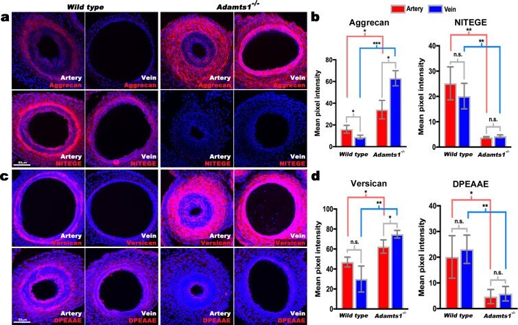
- Figure 6. Reduced aggrecan and versican proteolysis in Adamts1 -/- umbilical vessels. ( a,b ) E17.5 Adamts1 -/- umbilical vessels show increased aggrecan staining and reduced anti-NITEGE staining in ( a ), quantified in ( b ) (n = 3 cords each genotype, error bars indicate mean +-S.D. *, p
- Submitted by
- Invitrogen Antibodies (provider)
- Main image
- Experimental details
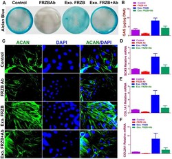
- Figure 2 Chondrogenic effects of FRZB on BMSCs in vitro . ( A ) Alcian blue staining of BMSCs in different treatment groups at 2 weeks to indicate GAG production in the culture plate. ( B ) Quantification of GAG production in generated cartilaginous tissues (n=6 for each). ( C ) Immunofluorescent assay of Aggrecan (green) expression and nucleus (blue) in different treatment groups observed under confocal microscopy. ( D - F ) Expression level of chondrogenic markers for BMSCs in different treatment groups (n=6 for each) in ( A ) (n=6 for each). *P < 0.05 between control group and other groups. Data are presented as averages +- SD. One-way analysis of variance (ANOVA) with post-hoc Tukey''s B test was applied. Ab, antibody; Exo, exogenous; ACAN , aggrecan; GAG, glycosaminoglycan.
- Submitted by
- Invitrogen Antibodies (provider)
- Main image
- Experimental details
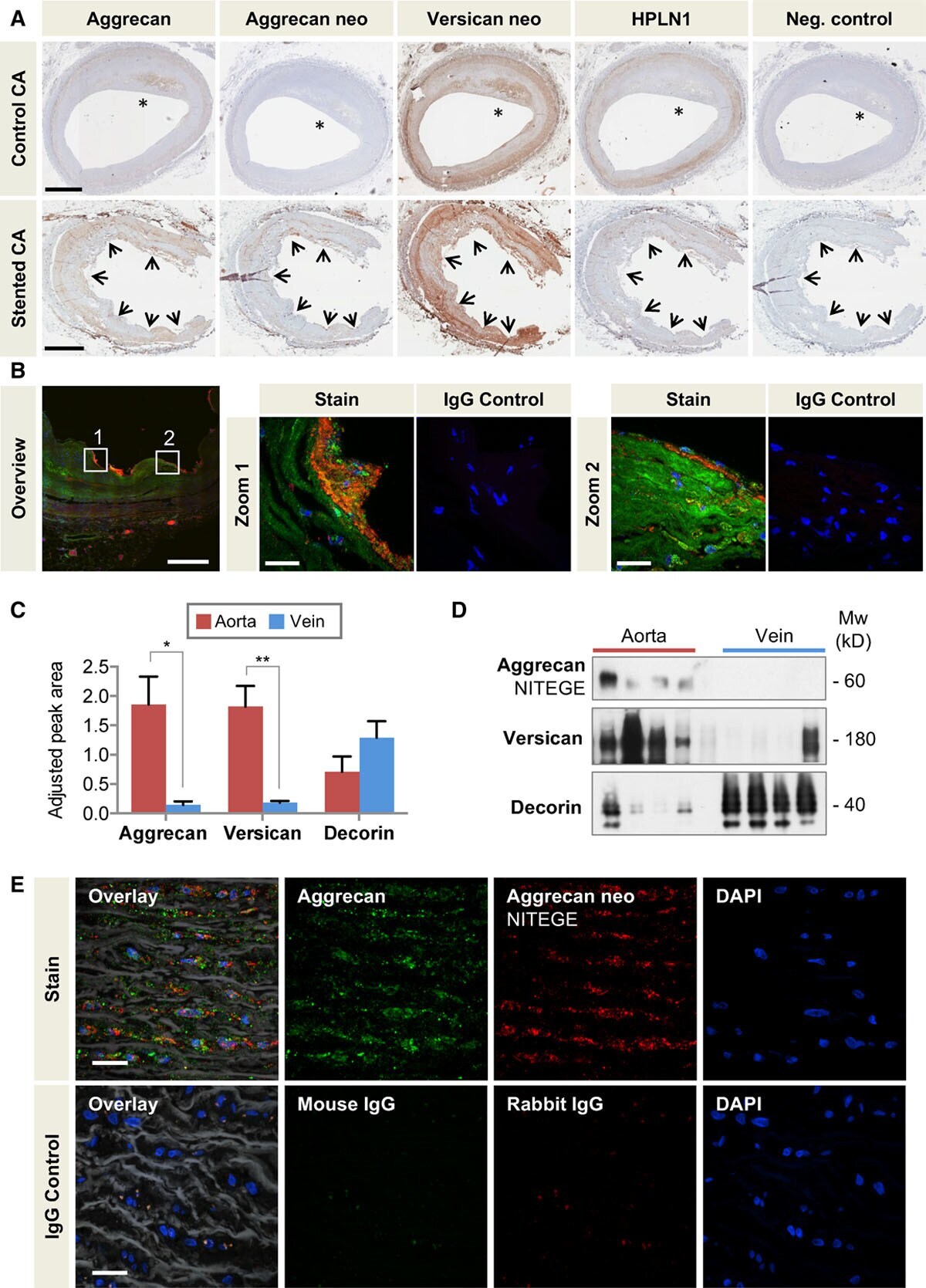
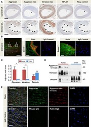
- Figure 6. Aggrecan in the human vasculature. A , Localization of aggrecan, the aggrecan NITEGE, and versican DPEAAE neoepitopes (neo), and HPLN1 in stented and control human coronary arteries with the presence of atherosclerosis (&ast). Arrows mark the contacts of the stent struts with coronary artery. Scale bars=1 mm. B , Colocalization of aggrecan (Alexa 633, displayed in green) and aggrecan NITEGE neoepitope (Alexa 568, displayed in red) in human stented coronary arteries by immunofluorescence. Note the presence of aggrecan fragments at the contacts of the stent struts with the artery and in the subendothelial layer (zoomed-in areas). Overview image 20x, scale bar=500 um; Zoomed-in areas 60x, scale bar=25 um. C , Adjusted peak area for aggrecan, versican, and decorin in the human thoracic aorta and saphenous veins as determined by targeted proteomics. n=7 per group. &ast P