Explore
 Validate
 Learn
Learn Western blot
 Immunoprecipitation
Antibody data
- Antibody Data
- Antigen structure
- References [4]
- Comments [0]
- Validations
- Western blot [1]
- Immunohistochemistry [1]
Submit
Validation data
Reference
Comment
Report error
- Product number
- AF5867 - Provider product page

- Provider
- Novus Biologicals
- Product name
- Sheep Polyclonal ADAMTS1 Antibody
- Antibody type
- Polyclonal
- Description
- Immunogen affinity purified. Detects human and mouse ADAMTS1 in direct ELISAs and Western blots. In direct ELISAs, less than 5% cross-reactivity with recombinant human (rh) ADAMTS4 and rhADAMTS15 is observed and less than 1% cross-reactivity with rhADAMTSL1.2, rhADAMTSL2, rhADAMTS5, rhADAMTS8, rhADAMTS10, rhADAMTS12, rhADAMTS13, and rhADAMTS16 is observed.
- Reactivity
- Human, Mouse
- Host
- Sheep
- Isotype
- IgG
- Vial size
- 100 ug
- Concentration
- LYOPH
- Storage
- Use a manual defrost freezer and avoid repeated freeze-thaw cycles. 12 months from date of receipt, -20 to -70 degreesC as supplied. 1 month, 2 to 8 degreesC under sterile conditions after reconstitution. 6 months, -20 to -70 degreesC under sterile conditions after reconstitution.
Submitted references ADAMTS-1 in abdominal aortic aneurysm.
A glucocorticoid- and diet-responsive pathway toggles adipocyte precursor cell activity in vivo.
Galnt1 is required for normal heart valve development and cardiac function.
ADAMTS1 inhibits lymphangiogenesis by attenuating phosphorylation of the lymphatic endothelial cell-specific VEGF receptor.
Vorkapic E, Folkesson M, Magnell K, Bohlooly-Y M, Länne T, Wågsäter D
PloS one 2017;12(6):e0178729
PloS one 2017;12(6):e0178729
A glucocorticoid- and diet-responsive pathway toggles adipocyte precursor cell activity in vivo.
Wong JC, Krueger KC, Costa MJ, Aggarwal A, Du H, McLaughlin TL, Feldman BJ
Science signaling 2016 Oct 25;9(451):ra103
Science signaling 2016 Oct 25;9(451):ra103
Galnt1 is required for normal heart valve development and cardiac function.
Tian E, Stevens SR, Guan Y, Springer DA, Anderson SA, Starost MF, Patel V, Ten Hagen KG, Tabak LA
PloS one 2015;10(1):e0115861
PloS one 2015;10(1):e0115861
ADAMTS1 inhibits lymphangiogenesis by attenuating phosphorylation of the lymphatic endothelial cell-specific VEGF receptor.
Inagaki J, Takahashi K, Ogawa H, Asano K, Faruk Hatipoglu O, Cilek MZ, Obika M, Ohtsuki T, Hofmann M, Kusachi S, Ninomiya Y, Hirohata S
Experimental cell research 2014 May 1;323(2):263-75
Experimental cell research 2014 May 1;323(2):263-75
No comments: Submit comment
Supportive validation
- Submitted by
- Novus Biologicals (provider)
- Main image

- Experimental details
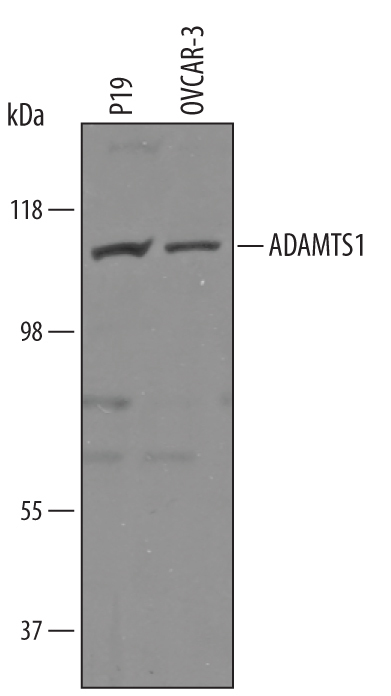
- Detection of Human and Mouse ADAMTS1 by Western Blot. Western blot shows lysates of P19 mouse embryonal carcinoma cell line and OVCAR-3 human ovarian carcinoma cell line. PVDF Membrane was probed with 1 µg/mL of Sheep Anti-Human/Mouse ADAMTS1 Antigen Affinity-purified Polyclonal Antibody (Catalog # AF5867) followed by HRP-conjugated Anti-Sheep IgG Secondary Antibody (Catalog # HAF016). A specific band was detected for ADAMTS1 at approximately 110 kDa (as indicated). This experiment was conducted under reducing conditions and using Immunoblot Buffer Group 8.
Supportive validation
- Submitted by
- Novus Biologicals (provider)
- Main image

- Experimental details
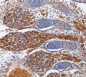
- ADAMTS1 in Mouse Embryo. ADAMTS1 was detected in immersion fixed frozen sections of mouse embryo (13 d.p.c.) using Sheep Anti-Human/Mouse ADAMTS1 Antigen Affinity-purified Polyclonal Antibody (Catalog # AF5867) at 15 µg/mL overnight at 4 °C. Tissue was stained using the Anti-Sheep HRP-DAB Cell & Tissue Staining Kit (brown; Catalog # CTS019) and counterstained with hematoxylin (blue). Specific staining was localized to cartilage primordium; dorsal ganglia cell bodies and processes. View our protocol for Chromogenic IHC Staining of Frozen Tissue Sections.