Explore
 Validate
 Learn
Learn Western blot
 Immunocytochemistry
Antibody data
- Antibody Data
- Antigen structure
- References [2]
- Comments [0]
- Validations
- Immunocytochemistry [4]
- Immunohistochemistry [2]
Submit
Validation data
Reference
Comment
Report error
- Product number
- MA1-22144 - Provider product page

- Provider
- Invitrogen Antibodies
- Product name
- Anti-Golgi protein 58k Monoclonal Antibody (58K-9)
- Antibody type
- Monoclonal
- Antigen
- Purifed from natural sources
- Description
- MA1-22146 detects Dynein intermediate chains in human, indian muntjac (deer), rat, chicken, and hamster samples. This antibody is know to detect cytoplasmic Dynein intermediate chains, and reactivity with the axonemal form is not known. MA1-22146 has been successfully used in ELISA, immunocytochemistry, immunofluorescence, immunoprecipitation and Western blot procedures. By Western blot, MA1-22146 detects a ~ 70 kDa triplet representing the Dynein intermediate chains. The MA1-22146 immunogen is cytoplasmic, full length, native, chicken dynein intermediate chain protein purified from chick brain.
- Reactivity
- Human, Rat, Bovine, Canine, Hamster, Porcine
- Host
- Mouse
- Isotype
- IgG
- Antibody clone number
- 58K-9
- Vial size
- 100 µL
- Concentration
- Lot Dependent
- Storage
- -20° C, Avoid Freeze/Thaw Cycles
Submitted references G protein βγ subunits regulate cardiomyocyte hypertrophy through a perinuclear Golgi phosphatidylinositol 4-phosphate hydrolysis pathway.
Mitochondrial dysfunction driven by the LRRK2-mediated pathway is associated with loss of Purkinje cells and motor coordination deficits in diabetic rat model.
Malik S, deRubio RG, Trembley M, Irannejad R, Wedegaertner PB, Smrcka AV
Molecular biology of the cell 2015 Mar 15;26(6):1188-98
Molecular biology of the cell 2015 Mar 15;26(6):1188-98
Mitochondrial dysfunction driven by the LRRK2-mediated pathway is associated with loss of Purkinje cells and motor coordination deficits in diabetic rat model.
Yang S, Xia C, Li S, Du L, Zhang L, Hu Y
Cell death & disease 2014 May 8;5:e1217
Cell death & disease 2014 May 8;5:e1217
No comments: Submit comment
Supportive validation
- Submitted by
- Invitrogen Antibodies (provider)
- Main image
- Experimental details
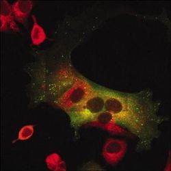
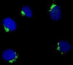
- Immunofluorescent analysis of Golgi protein, 58k in MCF-7 breast carcinoma cells expressing GFP fused CIN85 using a Golgi protein, 58k monoclonal antibody (Product # MA1-22144) at a dilution of 1:100.
- Submitted by
- Invitrogen Antibodies (provider)
- Main image
- Experimental details
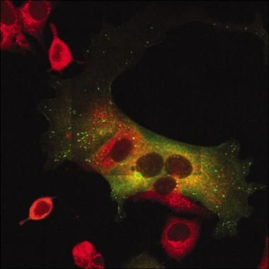
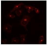
- Immunofluorescent analysis of Golgi protein, 58k in COS7 cells using a Golgi protein, 58k monoclonal antibody (Product # MA1-22144) at a dilution of 1:100 followed by hybridization with a secondary anti-mouse antibody (red) at a dilution of 1:100.
- Submitted by
- Invitrogen Antibodies (provider)
- Main image
- Experimental details
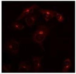
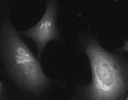
- Immunofluorescent analysis of Golgi protein, 58k using a Golgi protein, 58k monoclonal antibody (Product # MA1-22144). Cells were grown on coverslips, fixed with 70% methanol and permeabilized with 0.5% Triton X-100.

- Submitted by
- Invitrogen Antibodies (provider)
- Main image
- Experimental details
- Immunofluorescent analysis of Golgi protein in HeLa cells. Cells were fixed and permeabilized with 3% PFA plus 0.05% Triton X-100 in PBS and stained using a Golgi protein, 58k monoclonal antibody (Product # MA1-22144) at a dilution of 1:50, followed by detection using an anti-Mouse IgG conjugated to Cy3 at a dilution of 1:100.

Supportive validation
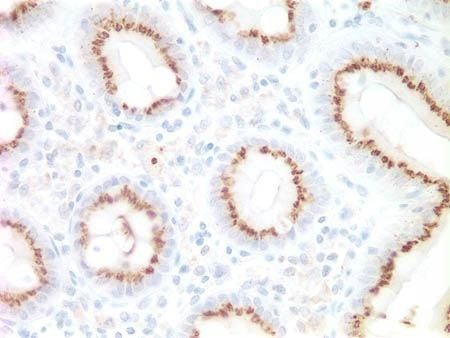
- Submitted by
- Invitrogen Antibodies (provider)
- Main image
- Experimental details
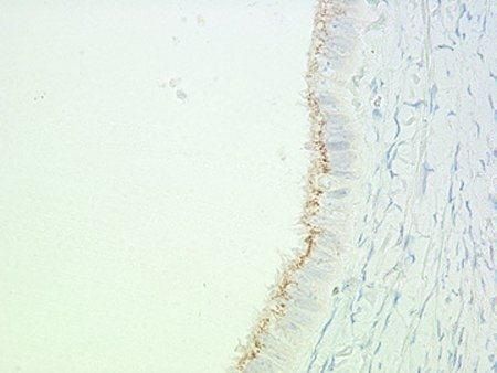
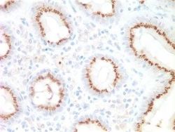
- Immunohistochemical analysis of Golgi protein, 58k in formalin-fixed, paraffin-embedded colon tissue using a Golgi protein, 58k monoclonal antibody (Product # MA1-22144) at a dilution of 1:1000 following heat-induced antigen retrieval.
- Submitted by
- Invitrogen Antibodies (provider)
- Main image

- Experimental details
- Immunohistochemical analysis of Golgi protein, 58k in formalin-fixed, paraffin-embedded ovary tissue using a Golgi protein, 58k monoclonal antibody (Product # MA1-22144) at a dilution of 1:1000 following heat-induced antigen retrieval.