 Explore
 Validate
 Learn
Learn Western blot
Antibody data
- Antibody Data
- Antigen structure
- References [0]
- Comments [0]
- Validations
- Western blot [3]
- Immunohistochemistry [1]
- Flow cytometry [1]
Submit
Validation data
Reference
Comment
Report error
- Product number
- PA1-9000 - Provider product page

- Provider
- Invitrogen Antibodies
- Product name
- Golgi protein 58k Polyclonal Antibody
- Antibody type
- Polyclonal
- Antigen
- Other
- Description
- This antibody is predicted to react with mouse, porcine and rat based on sequence homology. This antibody is tested in Peptide ELISA: antibody detection limit dilution 64,000.
- Reactivity
- Human, Mouse, Porcine
- Host
- Goat
- Isotype
- IgG
- Vial size
- 100 µg
- Concentration
- 0.5 mg/mL
- Storage
- -20° C, Avoid Freeze/Thaw Cycles
No comments: Submit comment
Supportive validation
- Submitted by
- Invitrogen Antibodies (provider)
- Main image

- Experimental details
- Western blot detection of 58K Golgi protein in human liver lysate using Product # PA1-9000.

- Submitted by
- Invitrogen Antibodies (provider)
- Main image

- Experimental details
- Western blot analysis of Golgi protein 58k by a Golgi protein 58k monoclonal antibody (Product # PA1-9000) at a concentration of 0.1 µg/mL. Human Liver lysate (35µg protein in RIPA buffer). Detected by chemiluminescence.

- Submitted by
- Invitrogen Antibodies (provider)
- Main image
- Experimental details
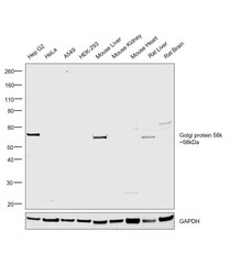
- Western blot was performed using Anti-Golgi protein 58k Polyclonal Antibody (Product # PA1-9000) and a 58kDa band corresponding to Golgi protein 58k was observed in Hep G2, Mouse Liver and Rat Liver but was not observed in HeLa, A549, HEK-293, Mouse Kidney, Mouse Heart and Rat Brain which are reported to be negative. Whole Cell Extract-WCL (30 µg lysate) of Hep G2 (Lane 1), HeLa (Lane 2), A549 (Lane 3), HEK-293 (Lane 4), Mouse Liver (Lane 5), Mouse Kidney (Lane 6), Mouse Heart (Lane 7), Rat Liver (Lane 8) and Rat Brain (Lane 9), were electrophoresed using NuPAGE™ 4-12% Bis-Tris Protein Gel (Product # NP0321BOX). Resolved proteins were then transferred onto a Nitrocellulose membrane (Product # IB23001) by iBlot® 2 Dry Blotting System (Product # IB21001). The blot was probed with the primary antibody (0.1ug/ml) and detected by chemiluminescence with Rabbit anti-Goat IgG (H+L) Superclonal™ Recombinant Secondary Antibody, HRP (Product # A27014, 1:4000 dilution) using the iBright FL 1000 (Product # A32752). Chemiluminescent detection was performed using Novex® ECL Chemiluminescent Substrate Reagent Kit (Product # WP20005).

Supportive validation
- Submitted by
- Invitrogen Antibodies (provider)
- Main image

- Experimental details
- Immunohistochemical analysis of Golgi protein 58k in Human Liver using a Golgi protein 58k monoclonal antibody (Product #PA1-9000) at 3.8 µg/mL. The Human Liver tissue section was paraffin embeded and detected using steamed antigen retrieval with citrate buffer pH 6, AP-staining.

Supportive validation
- Submitted by
- Invitrogen Antibodies (provider)
- Main image

- Experimental details
- Flow cytometric analysis of Golgi protein 58k in HepG2 cells using a polyclonal antibody (Product #PA1-9000). HepG2 cells (blue line) were paraformaldehyde fixed and permeabilized with 0.5% Triton. The primary antibody was incubated for one hour (10 µg/mL) followed by an Alexa Fluor 488 secondary antibody (1 µg/mL). IgG control: Unimmunized goat IgG (black line) followed by an Alexa Fluor 488 secondary antibody.
