 Explore
 Validate
 Learn
Learn Western blot
Antibody data
- Antibody Data
- Antigen structure
- References [0]
- Comments [0]
- Validations
- Western blot [1]
- ELISA [1]
- Immunoprecipitation [1]
- Immunohistochemistry [1]
Submit
Validation data
Reference
Comment
Report error
- Product number
- MA5-55658 - Provider product page

- Provider
- Invitrogen Antibodies
- Product name
- Golgi protein 58k Monoclonal Antibody (K92024_1D11)
- Antibody type
- Monoclonal
- Antigen
- Recombinant full-length protein
- Description
- Sequence of this protein is as follows: FSEGKNQEVI DAISGAITQT PGCVLLDVDA GPSTNRTVYT FVGPPECVVE GALNAARVAS RLIDMSRHQG EHPRMGALDV CPFIPVRGVS VDECVLCAQA FGQRLAEELD VPVYLYGEAA RMDSRRTLPA IRAGEYEALP KKLQQADWAP DFGPSSFVPS WGATATGARK FLIAFNINLL GTKEQAHRIA LNLREQGRGK DQPGRLKKVQ GIGWYLDEKN LAQVSTNLLD FEVTALHTVY EETCREAQEL SLPVVGSQLV GLVPLKALLD AAAFYCEKEN LFILEEEQRI RLVVSRLGLD SLCPFSPKER IIEYLVPERG
- Reactivity
- Human, Rat
- Host
- Mouse
- Isotype
- IgG
- Antibody clone number
- K92024_1D11
- Vial size
- 50 μg
- Concentration
- 1 mg/mL
- Storage
- Store at 4°C short term. For long term storage, store at -20°C, avoiding freeze/thaw cycles.
No comments: Submit comment
Supportive validation
- Submitted by
- Invitrogen Antibodies (provider)
- Main image
- Experimental details
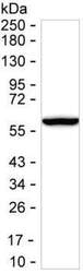
- Western blot analysis of Golgi protein 58k in 15 µg of rat liver tissue. Sample was run on 6-18% SDS-PAGE under reducing conditions, blotted onto nitrocellulose membrane, and peroxidase conjugated goat anti-mouse IgG was used as the secondary antibody. Golgi protein 58k band was visualized using ECL Substrate. Incubation with primary Golgi protein 58k monoclonal antibody (Product # MA5-55658) at a dilution of 1 µg/mL was used.
Supportive validation
- Submitted by
- Invitrogen Antibodies (provider)
- Main image
- Experimental details
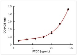
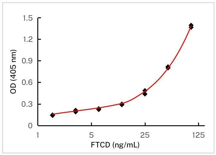
- ELISA using Golgi protein 58k as the antigen. Microtiter wells were coated with Golgi protein 58k monoclonal antibody (Product # MA5-55658) at a dilution of 3 µg/mL (capture). Peroxidase conjugated mouse anti-Golgi protein 58k monoclonal antibody was used as the detection antibody.
Supportive validation
- Submitted by
- Invitrogen Antibodies (provider)
- Main image
- Experimental details
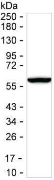
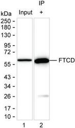
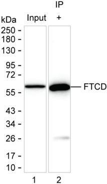
- Immunoprecipitation of Golgi protein 58k in 200 µg of rat liver tissue lysate. Samples are as follows: Lane 1: Rat liver tissue lysate, Lane 2: Golgi protein 58k immunoprecipitated from rat liver tissue lysate. After absorption with Protein G beads, the mixture was run on 6-18% SDS-PAGE, blotted onto nitrocellulose membrane, and peroxidase conjugated rabbit anti-mouse IgG (Light chain specific) was used as the secondary antibody. The isotype control antibody was KT82. Incubation of samples with Golgi protein 58k monoclonal antibody (Product # MA5-55658) at a dilution of 2.5 µg was used.
Supportive validation
- Submitted by
- Invitrogen Antibodies (provider)
- Main image
- Experimental details
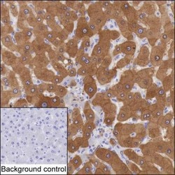
- Immunohistochemistry analysis of Golgi protein 58k in paraffin-embedded liver tissue. Sample was incubated with Golgi protein 58k monoclonal antibody (Product # MA5-55658) at a dilution of 2 µg/mL (RT, 1 hour). Antigen was retrieved through addition of boiling Tris/EDTA buffer pH 9 in a pressure cooker for 3 min. Endogenous peroxidase activity was quenched by incubating the sections with 3% H2O2 for 30 min at room temperature. Poly-peroxidase conjugated goat anti-mouse IgG was used as the secondary antibody. Diaminobenzidine was used as the chromogen. The section was counterstained with hematoxylin. A tissue section incubated with phosphate-buffered saline followed by incubation with the secondary antibody was used as the background control.
