 Explore
 Validate
 Learn
Learn Western blot
 Immunoprecipitation
Antibody data
- Antibody Data
- Antigen structure
- References [0]
- Comments [0]
- Validations
- Western blot [5]
- Immunocytochemistry [1]
Submit
Validation data
Reference
Comment
Report error
- Product number
- MA1-23284 - Provider product page

- Provider
- Invitrogen Antibodies
- Product name
- RPA2 Monoclonal Antibody (12F3.3)
- Antibody type
- Monoclonal
- Antigen
- Other
- Description
- Recommended positive controls: MCF7, T24, Neuro2A, GL261, BCL-1. Store product as a concentrated solution. Centrifuge briefly prior to opening the vial.
- Reactivity
- Human, Mouse
- Host
- Mouse
- Isotype
- IgG
- Antibody clone number
- 12F3.3
- Vial size
- 100 µL
- Concentration
- 1 mg/mL
- Storage
- Store at 4°C short term. For long term storage, store at -20°C, avoiding freeze/thaw cycles.
No comments: Submit comment
Supportive validation
- Submitted by
- Invitrogen Antibodies (provider)
- Main image

- Experimental details
- Western blot analysis of RPA32 in HeLa (IR treated) whole cell extract with no radiation (lane 1), 1hr (lane 2) 2hr (lane 3) and 4hr (lane 4) exposure using a RPA32 monoclonal antibody (Product # MA1-23284).

- Submitted by
- Invitrogen Antibodies (provider)
- Main image

- Experimental details
- RPA32 Polyclonal Antibody detects RPA32 protein by western blot analysis. Various whole cell extracts (30 µg) were separated by 12% SDS-PAGE, and blotted with RPA32 Polyclonal Antibody RPA2 Monoclonal Antibody (12F3.3) (Product # MA1-23284) diluted by 1:500. The HRP-conjugated anti-mouse IgG antibody was used to detect the primary antibody.

- Submitted by
- Invitrogen Antibodies (provider)
- Main image
- Experimental details
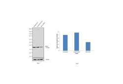
- Knockdown of RPA2 was achieved by transfecting HeLa with RPA2 specific siRNAs (Silencer® select Product # s12132, s12130). Western blot analysis (Fig. a) was performed using whole cell extracts (1% SDS) from the RPA2 knockdown cells (lane 3), non-specific scrambled siRNA transfected cells (lane 2) and untransfected cells (lane 1). The blots were probed with RPA2 Monoclonal Antibody (12F3.3) (Product # MA1-23284, 1 µg/mL) and Goat anti-Mouse IgG (H+L) Superclonal™ Secondary Antibody, HRP conjugate (Product # A28177, 0.25 µg/mL, 1:4000 dilution). Densitometric analysis of this western blot is shown in histogram (Fig. b). Decrease in signal upon siRNA mediated knock down confirms that antibody is specific to RPA2.

- Submitted by
- Invitrogen Antibodies (provider)
- Main image

- Experimental details
- Western blot analysis was performed on modified whole cell extracts (1%SDS) (30 µg lysate) of HeLa (Lane 1), MDA-MB-231 (Lane 2), PC-12 (Lane 3), Hep G2 (Lane 4), MCF7 (Lane 5), Jurkat (Lane 6), Neuro-2A (Lane 7), HEK293T (Lane 8) and NIH/3T3 (Lane 9). The blot was probed with Anti-RPA2 Monoclonal Antibody (12F3.3) (Product # MA1-23284, 1 µg/mL dilution) and detected by chemiluminescence using Goat anti-Mouse IgG (H+L) Superclonal™ Secondary Antibody, HRP conjugate (Product # A28177, 0.25 µg/mL, 1:4000 dilution). A 32 kDa band corresponding to RPA2 was observed across the cell lines tested.

- Submitted by
- Invitrogen Antibodies (provider)
- Main image

- Experimental details
- RPA32 Polyclonal Antibody detects RPA32 protein by western blot analysis. Various whole cell extracts (30 µg) were separated by 12% SDS-PAGE, and blotted with RPA32 Polyclonal Antibody RPA2 Monoclonal Antibody (12F3.3) (Product # MA1-23284) diluted by 1:500. The HRP-conjugated anti-mouse IgG antibody was used to detect the primary antibody.

Supportive validation
- Submitted by
- Invitrogen Antibodies (provider)
- Main image

- Experimental details
- RPA2 Monoclonal Antibody (12F3.3) detects RPA32 protein at DNA damage foci by immunofluorescent analysis. Sample: 2mM hydroxyurea treated (right) or untreated (left) HeLa cells were fixed in ice-cold MeOH for 5 min. Green: RPA32 protein stained by RPA2 Monoclonal Antibody (12F3.3) (Product # MA1-23284) diluted at 1:1,000. Blue: Hoechst 33342 staining. Scale bar = 10 µm.
