Explore
 Validate
 Learn
Learn Western blot
 Immunoprecipitation
Antibody data
- Antibody Data
- Antigen structure
- References [2]
- Comments [0]
- Validations
- Western blot [2]
- Immunocytochemistry [1]
Submit
Validation data
Reference
Comment
Report error
- Product number
- GTX70243 - Provider product page

- Provider
- GeneTex
- Proper citation
- GeneTex Cat#GTX70243, RRID:AB_372937
- Product name
- RPA32 antibody [12F3.3]
- Antibody type
- Monoclonal
- Reactivity
- Human, Mouse
- Host
- Mouse
Submitted references PARP3 affects the relative contribution of homologous recombination and nonhomologous end-joining pathways.
XRCC3 deficiency results in a defect in recombination and increased endoreduplication in human cells.
Beck C, Boehler C, Guirouilh Barbat J, Bonnet ME, Illuzzi G, Ronde P, Gauthier LR, Magroun N, Rajendran A, Lopez BS, Scully R, Boussin FD, Schreiber V, Dantzer F
Nucleic acids research 2014 May;42(9):5616-32
Nucleic acids research 2014 May;42(9):5616-32
XRCC3 deficiency results in a defect in recombination and increased endoreduplication in human cells.
Yoshihara T, Ishida M, Kinomura A, Katsura M, Tsuruga T, Tashiro S, Asahara T, Miyagawa K
The EMBO journal 2004 Feb 11;23(3):670-80
The EMBO journal 2004 Feb 11;23(3):670-80
No comments: Submit comment
Supportive validation
- Submitted by
- GeneTex (provider)
- Main image

- Experimental details
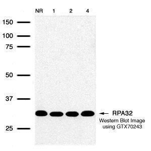
- Detection of human RPA32 protein using GeneTex RPA32 12F3.3 monoclonal antibody (GTX70243) in HeLa (IR treated) whole cell extract. No radiation, 1hr, 2hr, and 4hr exposure corresponding to lanes 1 through 4 respectively.
- Submitted by
- GeneTex (provider)
- Main image
- Experimental details
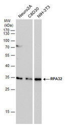
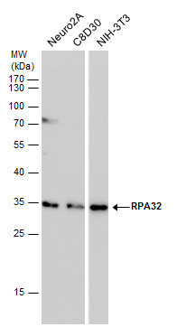
- RPA32 antibody detects RPA32 protein by western blot analysis. Various whole cell extracts (30 ?g) were separated by 12% SDS-PAGE, and blotted with RPA32 antibody (GTX70243) diluted by 1:500. The HRP-conjugated anti-mouse IgG antibody (GTX213111-01) was used to detect the primary antibody.
Supportive validation
- Submitted by
- GeneTex (provider)
- Main image

- Experimental details
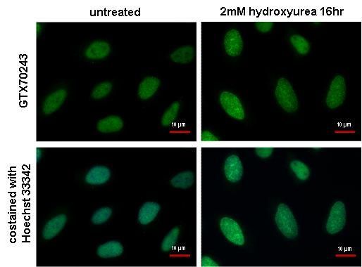
- RPA32 antibody [12F3.3] detects RPA32 protein at DNA damage foci by immunofluorescent analysis. Sample: 2mM hydroxyurea treated (right) or untreated (left) HeLa cells were fixed in ice-cold MeOH for 5 min. Green: RPA32 protein stained by RPA32 antibody [12F3.3] (GTX70243) diluted at 1:1000. Blue: Hoechst 33342 staining. Scale bar = 10 £gm.