Explore
 Validate
 Learn
Learn Western blot
Antibody data
- Antibody Data
- Antigen structure
- References [2]
- Comments [0]
- Validations
- Western blot [3]
- Immunocytochemistry [6]
- Immunohistochemistry [1]
- Other assay [1]
Submit
Validation data
Reference
Comment
Report error
- Product number
- PA5-22256 - Provider product page

- Provider
- Invitrogen Antibodies
- Product name
- RPA2 Polyclonal Antibody
- Antibody type
- Polyclonal
- Antigen
- Recombinant full-length protein
- Description
- Recommended positive controls: 293T, A431, BCL-1, PC-12, Rat2. Predicted reactivity: Mouse (90%), Rat (91%), Pig (93%), Chicken (81%), Rhesus Monkey (99%), Bovine (92%). Store product as a concentrated solution. Centrifuge briefly prior to opening the vial.
- Reactivity
- Human, Mouse, Rat
- Host
- Rabbit
- Isotype
- IgG
- Vial size
- 100 μL
- Concentration
- 1.64 mg/mL
- Storage
- Store at 4°C short term. For long term storage, store at -20°C, avoiding freeze/thaw cycles.
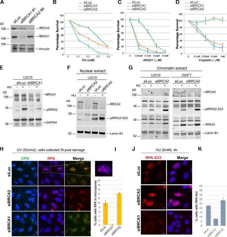
Submitted references E3 ligase RFWD3 is a novel modulator of stalled fork stability in BRCA2-deficient cells.
Glutaminase and poly(ADP-ribose) polymerase inhibitors suppress pyrimidine synthesis and VHL-deficient renal cancers.
Duan H, Mansour S, Reed R, Gillis MK, Parent B, Liu B, Sztupinszki Z, Birkbak N, Szallasi Z, Elia AEH, Garber JE, Pathania S
The Journal of cell biology 2020 Jun 1;219(6)
The Journal of cell biology 2020 Jun 1;219(6)
Glutaminase and poly(ADP-ribose) polymerase inhibitors suppress pyrimidine synthesis and VHL-deficient renal cancers.
Okazaki A, Gameiro PA, Christodoulou D, Laviollette L, Schneider M, Chaves F, Stemmer-Rachamimov A, Yazinski SA, Lee R, Stephanopoulos G, Zou L, Iliopoulos O
The Journal of clinical investigation 2017 May 1;127(5):1631-1645
The Journal of clinical investigation 2017 May 1;127(5):1631-1645
No comments: Submit comment
Supportive validation
- Submitted by
- Invitrogen Antibodies (provider)
- Main image
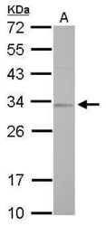
- Experimental details
- RPA2 Polyclonal Antibody detects RPA2 protein by Western blot analysis. A. 30 µg BCL-1 whole cell lysate/extract.12 % SDS-PAGE. RPA2 Polyclonal Antibody (Product # PA5-22256) dilution: 1:1,000.

- Submitted by
- Invitrogen Antibodies (provider)
- Main image

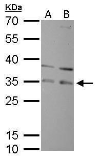
- Experimental details
- RPA2 Polyclonal Antibody detects RPA2 protein by Western blot analysis. A. 30 µg PC-12 whole cell lysate/extract. B. 30 µg Rat2 whole cell lysate/extract.12 % SDS-PAGE. RPA2 Polyclonal Antibody (Product # PA5-22256) dilution: 1:1,000.
- Submitted by
- Invitrogen Antibodies (provider)
- Main image
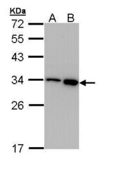
- Experimental details
- Western Blot using RPA2 Polyclonal Antibody (Product # PA5-22256). Sample (30 µg of whole cell lysate). Lane A: 293T. Lane B: A431. 12% SDS PAGE. RPA2 Polyclonal Antibody (Product # PA5-22256) diluted at 1:1,000.

Supportive validation
- Submitted by
- Invitrogen Antibodies (provider)
- Main image
- Experimental details
- Immunofluorescent analysis of RPA 32 kDa subunit in paraformaldehyde-fixed HeLa cells using a RPA 32 kDa subunit polyclonal antibody (Product # PA5-22256) at a 1:200 dilution.
- Submitted by
- Invitrogen Antibodies (provider)
- Main image

- Experimental details
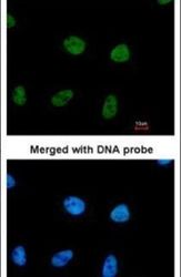
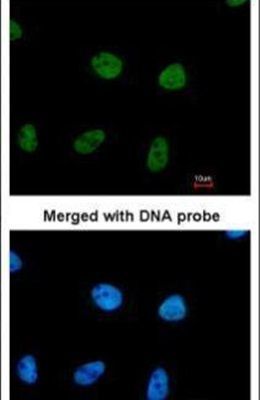
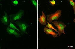
- RPA2 Polyclonal Antibody detects RPA32 protein at nucleus by immunofluorescent analysis. Sample: HeLa cells were fixed in 4% paraformaldehyde at RT for 15 min. Green: RPA32 stained by RPA2 Polyclonal Antibody (Product # PA5-22256) diluted at 1:500. Red: alpha Tubulin, a cytoskeleton marker, stained by alpha Tubulin antibody [GT114] (Product # MA5-31466) diluted at 1:1,000.

- Submitted by
- Invitrogen Antibodies (provider)
- Main image

- Experimental details
- RPA2 Polyclonal Antibody detects RPA32 protein at nucleus by immunofluorescent analysis. Sample: HeLa cells were fixed in 4% paraformaldehyde at RT for 15 min. Green: RPA32 stained by RPA2 Polyclonal Antibody (Product # PA5-22256) diluted at 1:500. Red: alpha Tubulin, a cytoskeleton marker, stained by alpha Tubulin antibody [GT114] (Product # MA5-31466) diluted at 1:1,000.

- Submitted by
- Invitrogen Antibodies (provider)
- Main image
- Experimental details
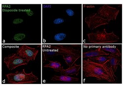
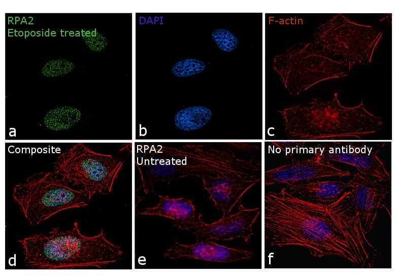
- Immunofluorescence analysis of RPA2 was performed using 70% confluent log phase HeLa cells treated with 30 µmol/L Etoposide for 1 hour. The cells were fixed with 4% paraformaldehyde for 10 minutes, permeabilized with 0.1% Triton™ X-100 for 10 minutes, and blocked with 1% BSA for 1 hour at room temperature. The cells were labeled with RPA2 Polyclonal Antibody (product # PA5-22256) at 5µg/mL in 0.1% BSA, incubated overnight at 4 degree Celsius and then labeled with Goat anti-Rabbit IgG (H+L) Superclonal™ Secondary Antibody, Alexa Fluor® 488 conjugate (Product # A27034) at a dilution of 1:2000 for 45 minutes at room temperature (Panel a: green). Nuclei (Panel b: blue) were stained with SlowFade® Gold Antifade Mountant with DAPI (Product # S36938). F-actin (Panel c: red) was stained with Rhodamine Phalloidin (Product # R415, 1:300). Panel d represents the merged image showing nuclear speckles upon Etoposide treatment. Panel e shows untreated cells without any staining. Panel f represents control cells with no primary antibody to assess background. The images were captured at 60X magnification.

- Submitted by
- Invitrogen Antibodies (provider)
- Main image

- Experimental details
- Immunofluorescence analysis of RPA2 was performed using 70% confluent log phase HeLa cells treated with 30 µmol/L Etoposide for 1 hour. The cells were fixed with 4% paraformaldehyde for 10 minutes, permeabilized with 0.1% Triton™ X-100 for 10 minutes, and blocked with 1% BSA for 1 hour at room temperature. The cells were labeled with RPA2 Polyclonal Antibody (product # PA5-22256) at 5µg/mL in 0.1% BSA, incubated overnight at 4 degree Celsius and then labeled with Goat anti-Rabbit IgG (Heavy Chain) Superclonal™ Secondary Antibody, Alexa Fluor® 488 conjugate (Product # A27034) at a dilution of 1:2000 for 45 minutes at room temperature (Panel a: green). Nuclei (Panel b: blue) were stained with SlowFade® Gold Antifade Mountant with DAPI (Product # S36938). F-actin (Panel c: red) was stained with Rhodamine Phalloidin (Product # R415, 1:300). Panel d represents the merged image showing nuclear speckles upon Etoposide treatment. Panel e shows untreated cells without any staining. Panel f represents control cells with no primary antibody to assess background. The images were captured at 60X magnification.
- Submitted by
- Invitrogen Antibodies (provider)
- Main image
- Experimental details
- RPA2 Polyclonal Antibody detects RPA32 protein at nucleus by immunofluorescent analysis. Sample: HeLa cells were fixed in 4% paraformaldehyde at RT for 15 min. Green: RPA32 stained by RPA2 Polyclonal Antibody (Product # PA5-22256) diluted at 1:500. Red: alpha Tubulin, a cytoskeleton marker, stained by alpha Tubulin antibody [GT114] (Product # MA5-31466) diluted at 1:1,000.

Supportive validation
- Submitted by
- Invitrogen Antibodies (provider)
- Main image
- Experimental details
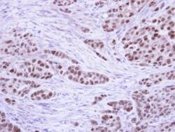
- Immunohistochemical analysis of paraffin-embedded A549 Xenograft, using RPA 32 kDa subunit (Product # PA5-22256) antibody at 1:100 dilution. Antigen Retrieval: EDTA based buffer, pH 8.0, 15 min.

Supportive validation
- Submitted by
- Invitrogen Antibodies (provider)
- Main image
- Experimental details
- NULL