 Explore
 Validate
 Learn
Learn Immunocytochemistry
Antibody data
- Antibody Data
- Antigen structure
- References [0]
- Comments [0]
- Validations
- Immunocytochemistry [1]
- Immunohistochemistry [1]
Submit
Validation data
Reference
Comment
Report error
- Product number
- MAB8654 - Provider product page

- Provider
- R&D Systems
- Product name
- Human Glut4 Antibody
- Antibody type
- Monoclonal
- Description
- Protein A or G purified from hybridoma culture supernatant. Detects human Glut4 in direct ELISAs.
- Reactivity
- Human
- Host
- Mouse
- Conjugate
- Unconjugated
- Antigen sequence
P14672- Isotype
- IgG
- Antibody clone number
- 925951
- Vial size
- 100 ug
- Storage
- Use a manual defrost freezer and avoid repeated freeze-thaw cycles. 12 months from date of receipt, -20 to -70 °C as supplied. 1 month, 2 to 8 °C under sterile conditions after reconstitution. 6 months, -20 to -70 °C under sterile conditions after reconstitution.
No comments: Submit comment
Supportive validation
- Submitted by
- R&D Systems (provider)
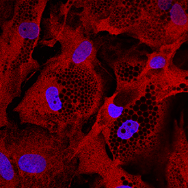
- Main image

- Experimental details
- Glut4 in Human Mesenchymal Stem Cells. Glut4 was detected in immersion fixed human mesenchymal stem cells differentiated to adipocytes using Mouse Anti-Human Glut4 Monoclonal Antibody (Catalog # MAB8654) at 10 µg/mL for 3 hours at room temperature. Cells were stained using the NorthernLights™ 557-conjugated Anti-Mouse IgG Secondary Antibody (red; Catalog # NL007) and counterstained with DAPI (blue). Specific staining was localized to cytoplasm. View our protocol for Fluorescent ICC Staining of Stem Cells on Coverslips.
Supportive validation
- Submitted by
- R&D Systems (provider)
- Main image
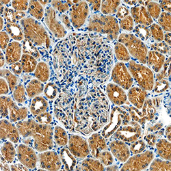
- Experimental details
- Glut4 in Human Kidney. Glut4 was detected in immersion fixed paraffin-embedded sections of human kidney using Mouse Anti-Human Glut4 Monoclonal Antibody (Catalog # MAB8654) at 15 µg/mL overnight at 4 °C. Tissue was stained using the Anti-Mouse HRP-DAB Cell & Tissue Staining Kit (brown; Catalog # CTS002) and counterstained with hematoxylin (blue). Specific staining was localized to cytoplasm in tubular epithelial cells. View our protocol for Chromogenic IHC Staining of Paraffin-embedded Tissue Sections.
