Explore
 Validate
 Learn
Learn Western blot
 Immunocytochemistry
Antibody data
- Antibody Data
- Antigen structure
- References [5]
- Comments [0]
- Validations
- Immunocytochemistry [3]
- Immunohistochemistry [1]
- Flow cytometry [2]
- Other assay [5]
Submit
Validation data
Reference
Comment
Report error
- Product number
- PA5-23052 - Provider product page

- Provider
- Invitrogen Antibodies
- Product name
- GLUT4 Polyclonal Antibody
- Antibody type
- Polyclonal
- Antigen
- Synthetic peptide
- Description
- This antibody is predicted to react with rat based on 100% sequence homology.
- Reactivity
- Human, Mouse, Rat
- Host
- Rabbit
- Isotype
- IgG
- Vial size
- 100 μL
- Concentration
- 1 mg/mL
- Storage
- -20°C or -80°C if preferred
Submitted references Diane-35 and Metformin Induce Autophagy and Apoptosis in Polycystic Ovary Syndrome Women with Early-Stage Endometrial Carcinoma.
Clenbuterol exerts antidiabetic activity through metabolic reprogramming of skeletal muscle cells.
CXCR1/2 Inhibitor Ladarixin Ameliorates the Insulin Resistance of 3T3-L1 Adipocytes by Inhibiting Inflammation and Improving Insulin Signaling.
Prior exercise in humans redistributes intramuscular GLUT4 and enhances insulin-stimulated sarcolemmal and endosomal GLUT4 translocation.
Photoperiodic modulation of thyroid hormone receptor (TR-α), deiodinase-2 (Dio-2) and glucose transporters (GLUT 1 and GLUT 4) expression in testis of adult golden hamster, Mesocricetus auratus.
Liu Y, Wang Y, Yao D, Chen X, Zhang F, Feng Y, Li X
Genes 2022 Jan 12;13(1)
Genes 2022 Jan 12;13(1)
Clenbuterol exerts antidiabetic activity through metabolic reprogramming of skeletal muscle cells.
Meister J, Bone DBJ, Knudsen JR, Barella LF, Velenosi TJ, Akhmedov D, Lee RJ, Cohen AH, Gavrilova O, Cui Y, Karsenty G, Chen M, Weinstein LS, Kleinert M, Berdeaux R, Jensen TE, Richter EA, Wess J
Nature communications 2022 Jan 10;13(1):22
Nature communications 2022 Jan 10;13(1):22
CXCR1/2 Inhibitor Ladarixin Ameliorates the Insulin Resistance of 3T3-L1 Adipocytes by Inhibiting Inflammation and Improving Insulin Signaling.
Castelli V, Brandolini L, d'Angelo M, Giorgio C, Alfonsetti M, Cocchiaro P, Lombardi F, Cimini A, Allegretti M
Cells 2021 Sep 6;10(9)
Cells 2021 Sep 6;10(9)
Prior exercise in humans redistributes intramuscular GLUT4 and enhances insulin-stimulated sarcolemmal and endosomal GLUT4 translocation.
Knudsen JR, Steenberg DE, Hingst JR, Hodgson LR, Henriquez-Olguin C, Li Z, Kiens B, Richter EA, Wojtaszewski JFP, Verkade P, Jensen TE
Molecular metabolism 2020 Sep;39:100998
Molecular metabolism 2020 Sep;39:100998
Photoperiodic modulation of thyroid hormone receptor (TR-α), deiodinase-2 (Dio-2) and glucose transporters (GLUT 1 and GLUT 4) expression in testis of adult golden hamster, Mesocricetus auratus.
Verma R, Haldar C
Journal of photochemistry and photobiology. B, Biology 2016 Dec;165:351-358
Journal of photochemistry and photobiology. B, Biology 2016 Dec;165:351-358
No comments: Submit comment
Supportive validation
- Submitted by
- Invitrogen Antibodies (provider)
- Main image

- Experimental details
- Immunofluorescent analysis of GLUT4 using a polyclonal antibody (Product # PA5-23052).
- Submitted by
- Invitrogen Antibodies (provider)
- Main image

- Experimental details
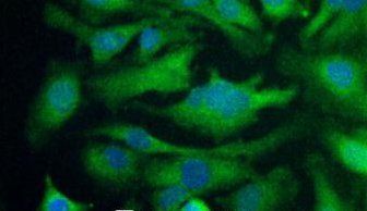
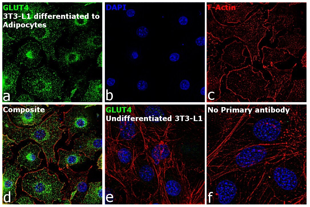
- Immunofluorescence analysis of GLUT4 was performed using 70% confluent log phase 3T3-L1 cells differentiated to adipocytes. The cells were fixed with 4% paraformaldehyde for 10 minutes, permeabilized with 0.1% Triton™ X-100 for 15 minutes, and blocked with 2% BSA for 1 hour at room temperature. The cells were labeled with GLUT4 Rabbit Polyclonal Antibody (Product # PA5-23052) at 5 µg/mL in 0.1% BSA, incubated at 4 degree celsius overnight and then labeled with Goat anti-Rabbit IgG (H+L) Superclonal™ Recombinant Secondary Antibody, Alexa Fluor® 488 (Product # A27034) at a dilution of 1:2000 for 45 minutes at room temperature (Panel a: green). Nuclei (Panel b: blue) were stained with ProLong™ Diamond Antifade Mountant with DAPI (Product # P36962). F-actin (Panel c: red) was stained with Rhodamine Phalloidin (Product # R415). Panel d represents the merged image showing cytoplasmic & membrane localization. Panel e represents 3T3-L1 (undifferentiated) cells having no expression of GLUT4. Panel f represents control cells with no primary antibody to assess background. The images were captured at 60X magnification.
- Submitted by
- Invitrogen Antibodies (provider)
- Main image

- Experimental details
- Immunofluorescence analysis of GLUT4 was performed using 70% confluent log phase 3T3-L1 cells differentiated to adipocytes. The cells were fixed with 4% paraformaldehyde for 10 minutes, permeabilized with 0.1% Triton™ X-100 for 15 minutes, and blocked with 2% BSA for 1 hour at room temperature. The cells were labeled with GLUT4 Rabbit Polyclonal Antibody (Product # PA5-23052) at 5 µg/mL in 0.1% BSA, incubated at 4 degree celsius overnight and then labeled with Goat anti-Rabbit IgG (Heavy Chain) Superclonal™ Recombinant Secondary Antibody, Alexa Fluor® 488 (Product # A27034) at a dilution of 1:2000 for 45 minutes at room temperature (Panel a: green). Nuclei (Panel b: blue) were stained with ProLong™ Diamond Antifade Mountant with DAPI (Product # P36962). F-actin (Panel c: red) was stained with Rhodamine Phalloidin (Product # R415). Panel d represents the merged image showing cytoplasmic & membrane localization. Panel e represents 3T3-L1 (undifferentiated) cells having no expression of GLUT4. Panel f represents control cells with no primary antibody to assess background. The images were captured at 60X magnification.

Supportive validation
- Submitted by
- Invitrogen Antibodies (provider)
- Main image

- Experimental details
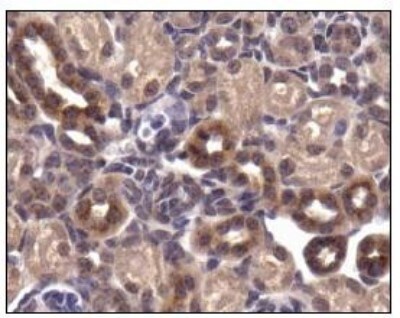
- Immunohistochemical analysis of GLUT4 in mouse kidney. Samples were incubated in GLUT4 polyclonal antibody (Product # PA5-23052).
Supportive validation
- Submitted by
- Invitrogen Antibodies (provider)
- Main image
- Experimental details
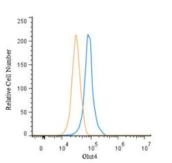
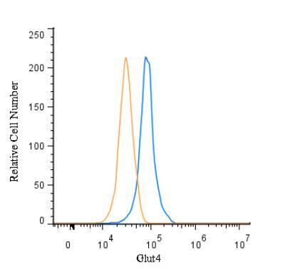
- Flow cytometry of GLUT4 in HepG2 and a matched isotype control. Samples were incubated in GLUT4 polyclonal antibody (Product # PA5-23052) using a dilution of 5 µg/mL for 30 minutes at room temperature followed by a Rabbit IgG (H+L) Cross-Adsorbed secondary antibody. Cells were fixed with 4% PFA and then permeabilized with 0.1% saponin.

- Submitted by
- Invitrogen Antibodies (provider)
- Main image
- Experimental details
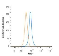
- Flow cytometry of GLUT4 in HepG2 and a matched isotype control. Samples were incubated in GLUT4 polyclonal antibody (Product # PA5-23052) using a dilution of 5 µg/mL for 30 minutes at room temperature followed by a Rabbit IgG (H+L) Cross-Adsorbed secondary antibody. Cells were fixed with 4% PFA and then permeabilized with 0.1% saponin.
Supportive validation
- Submitted by
- Invitrogen Antibodies (provider)
- Main image
- Experimental details
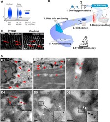
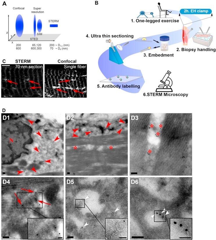
- Figure 1 Validation of a simple method for high-resolution imaging of endogenous GLUT4. (A) Theoretical resolvable volume for confocal microscopy, stimulated emission depletion (STED) and structured illumination super resolution microscopy (SIM) and the sample thinning enhanced resolution microscopy (STERM) used in the current study. D = diameter. It should be noted that these theoretical resolution values are estimates of the actual resolution of the techniques, since several factors affect the final imaging resolution. Figure modified from []. (B) Workflow overview for STERM. (1) Subjects underwent kicking exercise until exhaustion and (2) muscle biopsies were obtained and fixed before and after a hyperinsulinemic-euglycemic clamp. (3) Bundles of fibers were embedded in gelatin, sucrose infiltrated and mounted on cryo-pins. (4) Ultra-thin 70 nm sections were generated and (5) GLUT4 and subcellular compartment markers were labelled and (6) imaged. (C) Comparison of resolution by images obtained by confocal imaging of whole single mouse muscle fibers and 70 nm STERM sections prepared as illustrated in A. Red arrows mark vesicles which were better resolved by STERM. (D) Micrographs from transmission electron microscopy of human muscle showing GLUT4 in the perinuclear region (D1), the subsarcolemmal region (D2), the intrafibrillar region (D3), in tubule-vesicular regions (D4), in multi-vesicular endosomes (D5) and in individual vesicles (D6). Red arrowheads mark perinuclear and
- Submitted by
- Invitrogen Antibodies (provider)
- Main image

- Experimental details
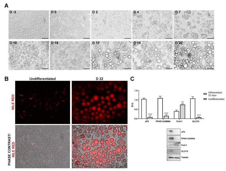
- Figure 1 ( A ) Contrast phase images at different time points showing the adipocyte differentiation (Scale bar = 100 mum), ( B ) Confluent 3T3-L1 cells stained with Nile Red prior to induction and after 22 days of differentiation. ( C ) Western blotting and relative densitometric analyses of 3T3-L1 prior differentiation and after 22 days of differentiation. Data are mean +- SE of 3 different experiments. t -test ** p < 0.005, *** p < 0.0005 vs. differentiated cells (N = 3).
- Submitted by
- Invitrogen Antibodies (provider)
- Main image
- Experimental details
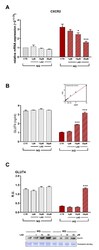
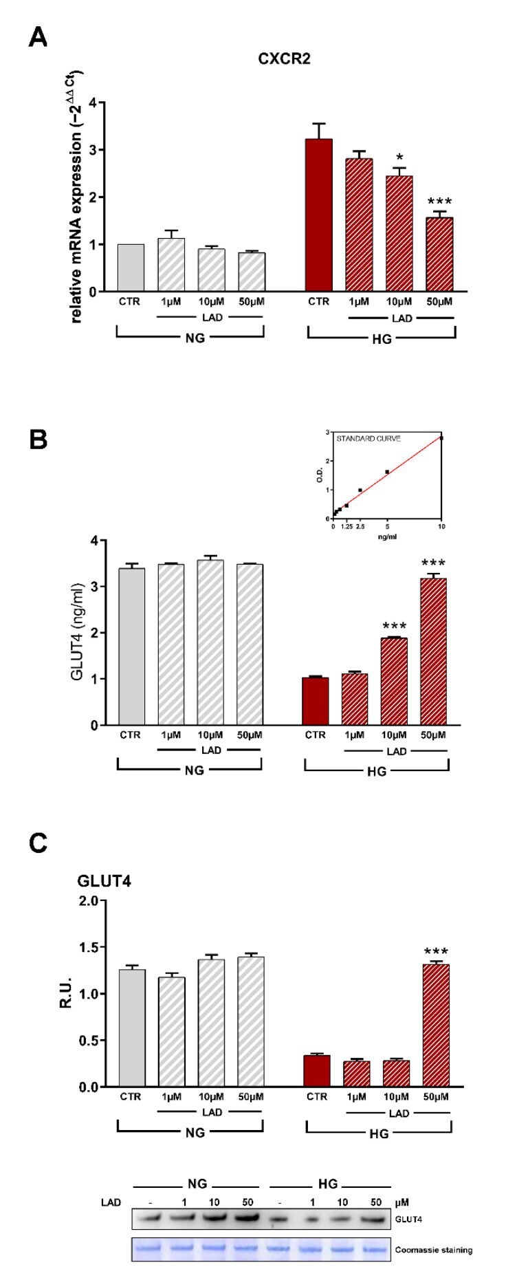
- Figure 2 ( A ) RT-PCR for CXCR2. Data are mean +- SE of 3 different experiments; One way ANOVA, * p < 0.05, *** p < 0.0005 vs. CTR (N = 3). ( B ) ELISA kit analyzing GLUT4 protein levels in plasma membrane and ( C ) Western blotting with relative densitometric analyses. A representative band is shown. Data are mean +- SE of 3 different experiments; One way ANOVA, *** p < 0.0005 vs. CTR (N = 3).
- Submitted by
- Invitrogen Antibodies (provider)
- Main image
- Experimental details
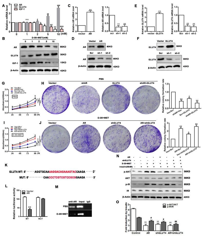
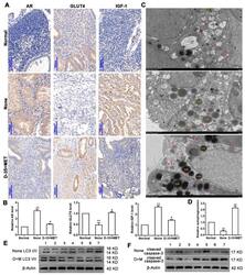
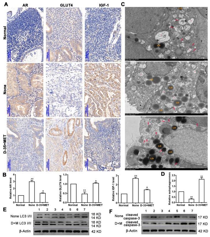
- Figure 1 Diane-35 and metformin reduced AR and IGF-1 expression and increased GLUT4 expression in the early-stage tumor tissue of PCOS patients with EC. Immunohistochemistry ( A ) and PCR analysis ( B ) for AR, GLUT4, and IGF-1 in endometrial carcinoma tissue, scale bars = 100 mum. Transmission electron microscope analysis of patients' tissues: autophagosomes: red arrow, autolysosome: white arrow, lysosome: red asterisk, and lipid droplet: LD, scale bars = 2 mum ( C , D ). Western blotting analysis of LC3 I/II ( E ) and cleaved caspase-3 ( F ) expression levels from 7 different patient samples. Data are shown as the mean +- S.D. * p < 0.05 and ** p < 0.01, ANOVA.
- Submitted by
- Invitrogen Antibodies (provider)
- Main image
- Experimental details
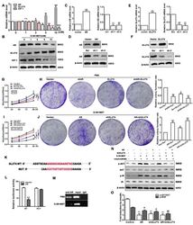
- Figure 4 Cell proliferation and insulin resistance is regulated by Diane-35 and metformin. Ishikawa cells were treated with increasing concentrations of Diane-35 and metformin for 24 h (0, 1, 2, 5, and 10 mM). Then, the expression of AR, GLUT4, and IGF-1 was assessed by qRT-PCR ( A ) and Western blotting ( B ). qRT-PCR ( C , E ) and Western blot ( D , F ) analyses were done for transfection efficiency measurements of the GLUT4 and AR expression vectors in Ishikawa cells. CCK-8 and colony formation assays were performed in transfected cells treated with ( G , H ) or without ( I , J ) Diane-35 and metformin (10 mM). ( K ) Wild and mutant promoter sequences of the GLUT4 gene. ( L ) Dual-luciferase reporting experiment for AR. ( M ) ChIP assay for binding validation between the AR and GLUT4 promoters. ( N , O ) Under Diane-35 and metformin treatment, p-AKT, AKT, p-IR, and IR expression levels in AR, and GLUT4-transfected Ishikawa cells with insulin stimulation. Data are shown as the mean +- S.D., n = 3. n.s. = no statistically significant differences. * p < 0.05, # p < 0.05, & p < 0.05, ** p < 0.01, ## p < 0.01, && p < 0.01 and *** p < 0.001, ### p < 0.001, &&& p < 0.001, ANOVA.