Explore
 Validate
 Learn
Learn Western blot
 ELISA
ELISAAntibody data
- Antibody Data
- Antigen structure
- References [0]
- Comments [0]
- Validations
- Western blot [1]
- Immunohistochemistry [1]
Submit
Validation data
Reference
Comment
Report error
- Product number
- PA1-32114 - Provider product page

- Provider
- Invitrogen Antibodies
- Product name
- p130 Polyclonal Antibody
- Antibody type
- Polyclonal
- Antigen
- Synthetic peptide
- Description
- Store as a concentrated solution. Centrifuge briefly prior to opening vial.
- Reactivity
- Human, Mouse, Rat
- Host
- Rabbit
- Isotype
- IgG
- Vial size
- 50 µL
- Concentration
- 85 mg/mL
- Storage
- Store at 4°C short term. For long term storage, store at -20°C, avoiding freeze/thaw cycles.
No comments: Submit comment
Supportive validation
- Submitted by
- Invitrogen Antibodies (provider)
- Main image

- Experimental details
- Western Blot of Rabbit Anti-Rb2 p130 Antibody. Lane 1: HEK 293 pcDNA3. Lane 2: HEK 293 pcDNA3-Rb2wt. Lane 3: HEK 293 pcDNA3-Rb2-PM19. Load: 30 µg per lane. Primary antibody: Anti-Rb2 antibody at 1:250 for overnight at 4°C. Secondary antibody: IRDye800™ rabbit secondary antibody at 1:10,000 for 45 min at RT. Block: 5% BLOTTO overnight at 4°C. Predicted/Observed size: 130 kDa for p130/Rb2.

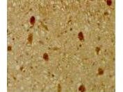
Supportive validation
- Submitted by
- Invitrogen Antibodies (provider)
- Main image

- Experimental details
- Immunohistochemical staining of mouse tissue using p130 Polyclonal Antibody (Product # PA1-32114). The staining shows the location of pRb2/p130 in developing mouse tissue. Other detection systems should yield similar results. Sections were cut at 5-7 µm, mounted on glass and dried overnight at 37&#deg;C. All sections were deparaffinized in xylene, rehydrated through a graded alcohol series and washed in phosphate-buffered saline (PBS). PBS was used for all subsequent washes and for antiserum dilution. Tissue sections were quenched sequentially in 0.5% hydrogen peroxide and blocked with diluted 10% normal goat anti-rabbit serum. Slides were incubated at 20º C for 1 h with rabbit anti-pRb2/p130 (1:500) dilution, washed, and then reacted with diluted goat anti-rabbit biotinylated antibody for 30 min. Slides were then reacted with streptavidin-peroxidase conjugate for 30 min at 20° C. Diaminobenzidine was used as the final chromogen. Negative controls for each tissue section were prepared by substituting the primary antiserum with pre-immune serum.