Explore
 Validate
 Learn
Learn Western blot
Antibody data
- Antibody Data
- Antigen structure
- References [3]
- Comments [0]
- Validations
- Western blot [4]
- Immunocytochemistry [2]
- Immunohistochemistry [2]
- Other assay [1]
Submit
Validation data
Reference
Comment
Report error
- Product number
- MA5-18288 - Provider product page

- Provider
- Invitrogen Antibodies
- Product name
- MCT1 Monoclonal Antibody (P14612)
- Antibody type
- Monoclonal
- Antigen
- Synthetic peptide
- Description
- Recommended positive controls: 293T. Store product as a concentrated solution. Centrifuge briefly prior to opening the vial.
- Reactivity
- Human
- Host
- Mouse
- Isotype
- IgG
- Antibody clone number
- P14612
- Vial size
- 100 μL
- Concentration
- 1 mg/mL
- Storage
- Store at 4°C short term. For long term storage, store at -20°C, avoiding freeze/thaw cycles.
Submitted references Using the Cancer Dependency Map to Identify the Mechanism of Action of a Cytotoxic Alkenyl Derivative from the Fruit of Choerospondias axillaris.
Bioinformatic analysis of membrane and associated proteins in murine cardiomyocytes and human myocardium.
Interfering cellular lactate homeostasis overcomes Taxol resistance of breast cancer cells through the microRNA-124-mediated lactate transporter (MCT1) inhibition.
Kil YS, Risinger AL, Petersen CL, Liang H, Grkovic T, O'Keefe BR, Mooberry SL, Cichewicz RH
Journal of natural products 2020 Mar 27;83(3):584-592
Journal of natural products 2020 Mar 27;83(3):584-592
Bioinformatic analysis of membrane and associated proteins in murine cardiomyocytes and human myocardium.
Lee SH, Hadipour-Lakmehsari S, Kim DH, Di Paola M, Kuzmanov U, Shah S, Lee JJ, Kislinger T, Sharma P, Oudit GY, Gramolini AO
Scientific data 2020 Dec 1;7(1):425
Scientific data 2020 Dec 1;7(1):425
Interfering cellular lactate homeostasis overcomes Taxol resistance of breast cancer cells through the microRNA-124-mediated lactate transporter (MCT1) inhibition.
Hou L, Zhao Y, Song GQ, Ma YH, Jin XH, Jin SL, Fang YH, Chen YC
Cancer cell international 2019;19:193
Cancer cell international 2019;19:193
No comments: Submit comment
Supportive validation
- Submitted by
- Invitrogen Antibodies (provider)
- Main image
- Experimental details
- Western Blot using MCT1 Monoclonal Antibody (P14612) (Product # MA5-18288). Unboiled various whole cell extracts (30 µg) were separated by 10% SDS-PAGE, and the membrane was blotted with MCT1 Monoclonal Antibody (P14612) (Product # MA5-18288) diluted at 1:1,000. The HRP-conjugated anti-mouse IgG antibody was used to detect the primary antibody, and the signal was developed with Trident ECL plus-Enhanced.
- Submitted by
- Invitrogen Antibodies (provider)
- Main image
- Experimental details
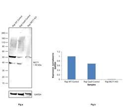
- Knockout of MCT1 was achieved by CRISPR-Cas9 genome editing using LentiArray™ Lentiviral sgRNA (Product # A32042, Assay ID CRISPR739575_LV) and LentiArray Cas9 Lentivirus (Product # A32064). Western blot analysis of MCT1 was performed by loading 50 µg of Raji Wild Type (Lane 1), Raji Cas9 (Lane 2) andRaji MCT1 KO (Lane 3) whole cell extracts. The samples were electrophoresed using NuPAGE™ Novex™ 4-12% Bis-Tris Protein Gel (Product # NP0322BOX). Resolved proteins were then transferred onto a nitrocellulose membrane (Product # IB23001) by iBlot® 2 Dry Blotting System (Product # IB21001). The blot was probed with Anti-MCT1 Monoclonal Antibody (P14612) (Product # MA5-18288, 1:1,000 dilution) and Goat anti-Mouse IgG (H+L) Superclonal™ Recombinant Secondary Antibody, HRP (Product # A28177, 1:5,000 dilution) using the iBright FL 1000 (Product # A32752). Chemiluminescent detection was performed using SuperSignal™ West Dura Extended Duration Substrate (Product # 34076). Loss of signal upon CRISPR mediated knockout (KO) using the LentiArray™ CRISPR product line confirms that antibody is specific to MCT1. An uncharacterized band was observed at ~50 kDa in all the samples.

- Submitted by
- Invitrogen Antibodies (provider)
- Main image
- Experimental details
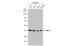
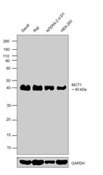
- Western blot was performed using Anti-MCT1 Monoclonal Antibody (P14612) (Product # MA5-18288) and a 40 kDa band corresponding to MCT1 was observed across the cell lines tested. Whole cell extracts (30 µg lysate) of Daudi (Lane 1), Raji (Lane 2), NTERA-2 cl.D1 (Lane 3) and HEK-293 (Lane 4) were electrophoresed using Novex® NuPAGE™ 10% Bis-Tris Protein Gel (Product # NP0302BOX). Resolved proteins were then transferred onto a nitrocellulose membrane (Product # IB23001) by iBlot® 2 Dry Blotting System (Product # IB21001). The blot was probed with the primary antibody (1:1000 dilution) and detected by chemiluminescence with Goat anti-Mouse IgG (H+L), Superclonal™ Recombinant Secondary Antibody, HRP (Product # A28177, 1:4000 dilution) using the iBright FL 1000 (Product # A32752), using the iBright FL 1000 (Product # A32752). Chemiluminescent detection was performed using Novex® ECL Chemiluminescent Substrate Reagent Kit (Product # WP20005).
- Submitted by
- Invitrogen Antibodies (provider)
- Main image
- Experimental details
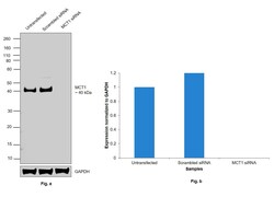
- Knockdown of MCT1 was achieved by transfecting HEK-293 with MCT1 specific siRNAs (Silencer® select Product # s580). Western blot analysis (Fig. a) was performed using whole cell extracts from the MCT1 knockdown cells (Lane 3), non-specific scrambled siRNA transfected cells (Lane 2) and untransfected cells (Lane 1). The blot was probed with MCT1 Monoclonal Antibody (P14612) (Product # MA5-18288, 1:1000 dilution) and Goat anti-Mouse IgG (H+L), Superclonal™ Recombinant Secondary Antibody, HRP (Product # A28177, 1:4000 dilution) using the iBright FL 1000 (Product # A32752). Densitometric analysis of this Western Blot is shown in histogram (Fig. b). Decrease in signal upon siRNA mediated knock down confirms that antibody is specific to MCT1.

Supportive validation
- Submitted by
- Invitrogen Antibodies (provider)
- Main image

- Experimental details
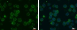
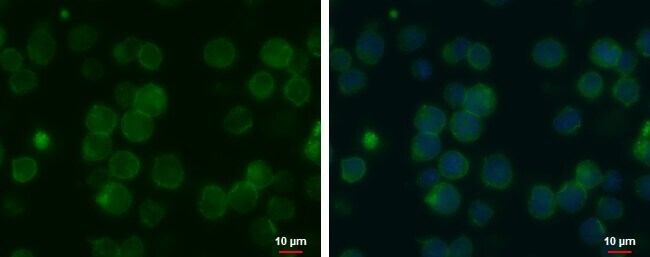
- MCT1 Monoclonal Antibody (P14612) detects MCT1 protein at cell membrane by immunofluorescent analysis. Sample: THP-1 cells were fixed in 4% paraformaldehyde at RT for 15 min. Green: MCT1 protein stained by MCT1 Monoclonal Antibody (P14612) (Product # MA5-18288) diluted at 1:500. Blue: Hoechst 33342 staining. Scale bar = 10 µm.

- Submitted by
- Invitrogen Antibodies (provider)
- Main image
- Experimental details
- MCT1 Monoclonal Antibody (P14612) detects MCT1 protein at cell membrane by immunofluorescent analysis. Sample: THP-1 cells were fixed in 4% paraformaldehyde at RT for 15 min. Green: MCT1 protein stained by MCT1 Monoclonal Antibody (P14612) (Product # MA5-18288) diluted at 1:500. Blue: Hoechst 33342 staining. Scale bar = 10 µm.
Supportive validation
- Submitted by
- Invitrogen Antibodies (provider)
- Main image

- Experimental details
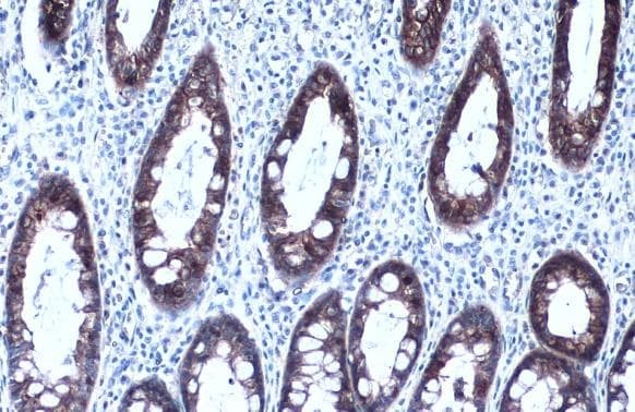
- MCT1 Monoclonal Antibody (P14612) detects MCT1 protein at cell membrane by immunohistochemical analysis. Sample: Paraffin-embedded human colon. MCT1 stained by MCT1 Monoclonal Antibody (P14612) (Product # MA5-18288) diluted at 1:500. Antigen Retrieval: Citrate buffer, pH 6.0, 15 min.

- Submitted by
- Invitrogen Antibodies (provider)
- Main image

- Experimental details
- MCT1 Monoclonal Antibody (P14612) detects MCT1 protein at cell membrane by immunohistochemical analysis. Sample: Paraffin-embedded human colon. MCT1 stained by MCT1 Monoclonal Antibody (P14612) (Product # MA5-18288) diluted at 1:500. Antigen Retrieval: Citrate buffer, pH 6.0, 15 min.
Supportive validation
- Submitted by
- Invitrogen Antibodies (provider)
- Main image
- Experimental details
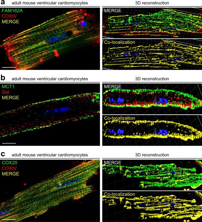
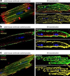
- Fig. 8 Co-immunofluorescence analysis demonstrates colocalization of FAM162A and COX20 with known mitochondrial marker, COXIV, and MCT1 colocalization with known plasma membrane protein, Galphai, in isolated adult mouse cardiomyocytes. ( a ) Immunofluorescence analysis of FAM162A (green) co-stained with mitochondrial protein, COXIV (red) in acutely isolated adult mouse cardiomyocytes. Three-dimensional reconstructive analysis demonstrates regions of colocalization (yellow) with a Pearson's coefficient p > 0.5. Scale, 10 mum. ( b ) Immunofluorescence analysis of MCT1 (green) co-stained with known plasma membrane protein, Galphai (red) in acutely isolated adult mouse cardiomyocytes. Three-dimensional reconstructive analysis demonstrates regions of colocalization (yellow) with a Pearson's coefficient p > 0.5. Scale, 10mum. ( c ) Immunofluorescence analysis of COX20 (green) co-stained with mitochondrial protein, COXIV (red) in acutely isolated adult mouse cardiomyocytes. Three-dimensional reconstructive analysis demonstrates regions of colocalization (yellow) with a Pearson's coefficient p > 0.5. Scale, 10mum. Nuclear staining was visualized with Hoechst staining (blue). All images shown are representative of approximately 30-40 total images captured per condition, n = 3 independent biological replicates. All original uncropped microscopy images were uploaded to figshare (10.6084/m9.figshare.11844972.v12).