 Explore
 Validate
 Learn
Learn Western blot
Antibody data
- Antibody Data
- Antigen structure
- References [1]
- Comments [0]
- Validations
- Western blot [2]
- Immunohistochemistry [1]
- Other assay [1]
Submit
Validation data
Reference
Comment
Report error
- Product number
- PA5-72957 - Provider product page

- Provider
- Invitrogen Antibodies
- Product name
- MCT1 Polyclonal Antibody
- Antibody type
- Polyclonal
- Antigen
- Synthetic peptide
- Reactivity
- Human, Mouse, Rat
- Host
- Rabbit
- Isotype
- IgG
- Vial size
- 100 µg
- Concentration
- 1 mg/mL
- Storage
- Store at 4°C short term. For long term storage, store at -20°C, avoiding freeze/thaw cycles.
Submitted references Bioinformatic analysis of membrane and associated proteins in murine cardiomyocytes and human myocardium.
Lee SH, Hadipour-Lakmehsari S, Kim DH, Di Paola M, Kuzmanov U, Shah S, Lee JJ, Kislinger T, Sharma P, Oudit GY, Gramolini AO
Scientific data 2020 Dec 1;7(1):425
Scientific data 2020 Dec 1;7(1):425
No comments: Submit comment
Supportive validation
- Submitted by
- Invitrogen Antibodies (provider)
- Main image

- Experimental details
- Western blot analysis of MCT1 in Jurkat cell lysate. Samples were incubated in MCT1 polyclonal antibody (Product # PA5-72957) using a dilution of 2.5 µg/mL. Jurkat Tag cells were transfected with the expression vector (vector) or with the Foxp3 expression construct (Foxp3). 24 hours after transfection, cells were lysed and analyzed by Western blot with anti-Foxp3 antiserum (1:2000 dilution).

- Submitted by
- Invitrogen Antibodies (provider)
- Main image

- Experimental details
- Western blot was performed using Anti-MCT1 Polyclonal Antibody (Product # PA5-72957) and a 40 kDa band corresponding to MCT1 was observed across the tissues tested except in Mouse Brain and Rat Brain which are reported to be negative. Tissue extracts (30 µg lysate) of Mouse Adipose (Lane 1), Rat Adipose (Lane 2), Mouse Brown Fat (Lane 3), Rat Brown Fat (Lane 4), Mouse Kidney (Lane 5), Mouse Lung (Lane 6), Mouse Liver (Lane 7), Mouse Skeletal Muscle (Lane 8), Mouse Brain (Lane 9) and Rat Brain (Lane 10) were electrophoresed using Novex® NuPAGE™ 10% Bis-Tris Protein Gel (Product # NP0302BOX). Resolved proteins were then transferred onto a nitrocellulose membrane (Product # IB23001) by iBlot® 2 Dry Blotting System (Product # IB21001). The blot was probed with the primary antibody (2 µg/mL) and detected by chemiluminescence with Goat anti-Rabbit IgG (H+L), Superclonal™ Recombinant Secondary Antibody, HRP (Product # A27036, 1:4000 dilution), using the iBright FL 1000 (Product # A32752). Chemiluminescent detection was performed using Novex® ECL Chemiluminescent Substrate Reagent Kit (Product # WP20005).

Supportive validation
- Submitted by
- Invitrogen Antibodies (provider)
- Main image

- Experimental details
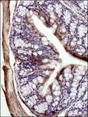
- Immunohistochemical analysis of MCT1 in the submucosa of mouse intestine. Samples were incubated in MCT1 polyclonal antibody (Product # PA5-72957). Strong staining of lumen and crypt cells was observed with weaker cytoplasmic staining observed in the submucosa of mouse intestine.
Supportive validation
- Submitted by
- Invitrogen Antibodies (provider)
- Main image

- Experimental details
- Fig. 8 Co-immunofluorescence analysis demonstrates colocalization of FAM162A and COX20 with known mitochondrial marker, COXIV, and MCT1 colocalization with known plasma membrane protein, Galphai, in isolated adult mouse cardiomyocytes. ( a ) Immunofluorescence analysis of FAM162A (green) co-stained with mitochondrial protein, COXIV (red) in acutely isolated adult mouse cardiomyocytes. Three-dimensional reconstructive analysis demonstrates regions of colocalization (yellow) with a Pearson's coefficient p > 0.5. Scale, 10 mum. ( b ) Immunofluorescence analysis of MCT1 (green) co-stained with known plasma membrane protein, Galphai (red) in acutely isolated adult mouse cardiomyocytes. Three-dimensional reconstructive analysis demonstrates regions of colocalization (yellow) with a Pearson's coefficient p > 0.5. Scale, 10mum. ( c ) Immunofluorescence analysis of COX20 (green) co-stained with mitochondrial protein, COXIV (red) in acutely isolated adult mouse cardiomyocytes. Three-dimensional reconstructive analysis demonstrates regions of colocalization (yellow) with a Pearson's coefficient p > 0.5. Scale, 10mum. Nuclear staining was visualized with Hoechst staining (blue). All images shown are representative of approximately 30-40 total images captured per condition, n = 3 independent biological replicates. All original uncropped microscopy images were uploaded to figshare (10.6084/m9.figshare.11844972.v12).
