 Explore
 Validate
 Learn
Learn Western blot
Antibody data
- Antibody Data
- Antigen structure
- References [0]
- Comments [0]
- Validations
- Western blot [5]
- Immunohistochemistry [1]
- Flow cytometry [1]
Submit
Validation data
Reference
Comment
Report error
- Product number
- PA5-111847 - Provider product page

- Provider
- Invitrogen Antibodies
- Product name
- MCT1 (extracellular) Polyclonal Antibody
- Antibody type
- Polyclonal
- Antigen
- Synthetic peptide
- Description
- Applications Reported: This SJ25C1 antibody has been reported for use in flow cytometric analysis.
- Reactivity
- Human, Mouse, Rat
- Host
- Rabbit
- Isotype
- IgG
- Vial size
- 50 µL
- Concentration
- 0.8 mg/mL
- Storage
- -20°C
No comments: Submit comment
Supportive validation
- Submitted by
- Invitrogen Antibodies (provider)
- Main image
- Experimental details
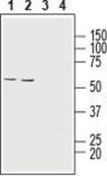
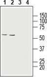
- Western Blot analysis of MCT1 was performed in human HT-29 colon adenocarcinoma cell line lysate (lanes 1 and 3) and human ARPE-19 retinal pigment epithelium cell line lysate (lanes 2 and 4). Lane 1,2: MCT1 (extracellular) Antibody (Product # PA5-111847) at a dilution of 1:200. Lane 3,4: MCT1 (extracellular) Antibody preincubated with the negative control antigen.

- Submitted by
- Invitrogen Antibodies (provider)
- Main image
- Experimental details
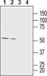
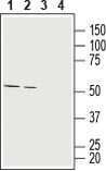
- Western Blot analysis of MCT1 was performed in rat (lanes 1 and 3) and mouse (lanes 2 and 4) brain lysates. Lane 1,2: MCT1 (extracellular) Antibody (Product # PA5-111847) at a dilution of 1:200. Lane 3,4: MCT1 (extracellular) Antibody preincubated with the negative control antigen.
- Submitted by
- Invitrogen Antibodies (provider)
- Main image
- Experimental details
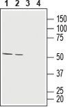
- Western Blot analysis of MCT1 was performed in human HT-29 colon adenocarcinoma cell line lysate (lanes 1 and 3) and human ARPE-19 retinal pigment epithelium cell line lysate (lanes 2 and 4). Lane 1,2: MCT1 (extracellular) Antibody (Product # PA5-111847) at a dilution of 1:200. Lane 3,4: MCT1 (extracellular) Antibody preincubated with the negative control antigen.
- Submitted by
- Invitrogen Antibodies (provider)
- Main image

- Experimental details
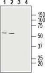
- Western Blot analysis of MCT1 was performed in rat (lanes 1 and 3) and mouse (lanes 2 and 4) brain lysates. Lane 1,2: MCT1 (extracellular) Antibody (Product # PA5-111847) at a dilution of 1:200. Lane 3,4: MCT1 (extracellular) Antibody preincubated with the negative control antigen.
- Submitted by
- Invitrogen Antibodies (provider)
- Main image
- Experimental details
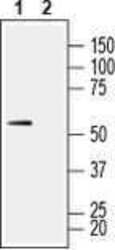
- Western Blot analysis of MCT1 was performed in rat skeletal muscle lysate. Lane 1: MCT1 (extracellular) Antibody (Product # PA5-111847) at a dilution of 1:200. Lane 2: MCT1 (extracellular) Antibody preincubated with the negative control antigen.

Supportive validation
- Submitted by
- Invitrogen Antibodies (provider)
- Main image

- Experimental details
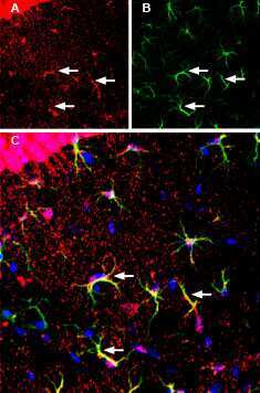
- Immunohistochemistry analysis of MCT1 in immersion-fixed, frozen mouse hippocampus tissue sections using MCT1 (extracellular) Antibody (Product # PA5-111847) at a dilution of 1:200, followed by goat-anti-rabbit-Cy3. A) MCT1 staining (red) in CA1 hippocampal region, is detected in the pyramidal layer (P) and in small cell outlines (arrows). B) GFAP staining (green) is observed in astrocyte outlines (arrows). C) Merge of the two images demonstrates co-localization of MCT1 and GFAP in astrocytes (arrows). Cell nuclei are stained with DAPI (blue).
Supportive validation
- Submitted by
- Invitrogen Antibodies (provider)
- Main image

- Experimental details
- Flow Cytometry analysis of MCT1 in live intact human Jurkat T-cell leukemia cells. Black: Cells. Pink: Cells and goat-anti-rabbit-FITC. Green: Cells, MCT1 (extracellular) Antibody (Product # PA5-111847), and goat-anti-rabbit-FITC.
