Explore
 Validate
 Learn
Learn Western blot
 Immunoprecipitation
Antibody data
- Antibody Data
- Antigen structure
- References [3]
- Comments [0]
- Validations
- Western blot [1]
- Immunocytochemistry [1]
Submit
Validation data
Reference
Comment
Report error
- Product number
- AF4119 - Provider product page

- Provider
- R&D Systems
- Product name
- Human alpha-L-Iduronidase/IDUA Antibody
- Antibody type
- Polyclonal
- Description
- Antigen Affinity-purified. Detects human alpha-L-Iduronidase/IDUA in direct ELISAs and Western blots.
- Reactivity
- Human
- Host
- Sheep
- Conjugate
- Unconjugated
- Antigen sequence
AAA81589- Isotype
- IgG
- Vial size
- 100 ug
- Concentration
- LYOPH
- Storage
- Use a manual defrost freezer and avoid repeated freeze-thaw cycles. 12 months from date of receipt, -20 to -70 °C as supplied. 1 month, 2 to 8 °C under sterile conditions after reconstitution. 6 months, -20 to -70 °C under sterile conditions after reconstitution.
Submitted references ZFN-Mediated In Vivo Genome Editing Corrects Murine Hurler Syndrome.
Toxicology Study of Intra-Cisterna Magna Adeno-Associated Virus 9 Expressing Human Alpha-L-Iduronidase in Rhesus Macaques.
Mutation in VPS33A affects metabolism of glycosaminoglycans: a new type of mucopolysaccharidosis with severe systemic symptoms.
Ou L, DeKelver RC, Rohde M, Tom S, Radeke R, St Martin SJ, Santiago Y, Sproul S, Przybilla MJ, Koniar BL, Podetz-Pedersen KM, Laoharawee K, Cooksley RD, Meyer KE, Holmes MC, McIvor RS, Wechsler T, Whitley CB
Molecular therapy : the journal of the American Society of Gene Therapy 2019 Jan 2;27(1):178-187
Molecular therapy : the journal of the American Society of Gene Therapy 2019 Jan 2;27(1):178-187
Toxicology Study of Intra-Cisterna Magna Adeno-Associated Virus 9 Expressing Human Alpha-L-Iduronidase in Rhesus Macaques.
Hordeaux J, Hinderer C, Goode T, Katz N, Buza EL, Bell P, Calcedo R, Richman LK, Wilson JM
Molecular therapy. Methods & clinical development 2018 Sep 21;10:79-88
Molecular therapy. Methods & clinical development 2018 Sep 21;10:79-88
Mutation in VPS33A affects metabolism of glycosaminoglycans: a new type of mucopolysaccharidosis with severe systemic symptoms.
Kondo H, Maksimova N, Otomo T, Kato H, Imai A, Asano Y, Kobayashi K, Nojima S, Nakaya A, Hamada Y, Irahara K, Gurinova E, Sukhomyasova A, Nogovicina A, Savvina M, Yoshimori T, Ozono K, Sakai N
Human molecular genetics 2017 Jan 1;26(1):173-183
Human molecular genetics 2017 Jan 1;26(1):173-183
No comments: Submit comment
Supportive validation
- Submitted by
- R&D Systems (provider)
- Main image
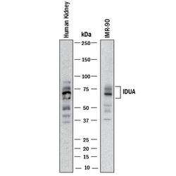
- Experimental details
- Detection of Human alpha-L-Iduronidase/IDUA by Western Blot. Western blot shows lysates of human kidney tissue and IMR-90 human lung fibroblast cell line. PVDF membrane was probed with 1 µg/mL of Sheep Anti-Human alpha-L-Iduronidase/IDUA Antigen Affinity-purified Polyclonal Antibody (Catalog # AF4119) followed by HRP-conjugated Anti-Sheep IgG Secondary Antibody (Catalog # HAF016). Specific bands were detected for alpha-L-Iduronidase/IDUA at approximately 74 kDa (as indicated). This experiment was conducted under reducing conditions and using Immunoblot Buffer Group 1.

Supportive validation
- Submitted by
- R&D Systems (provider)
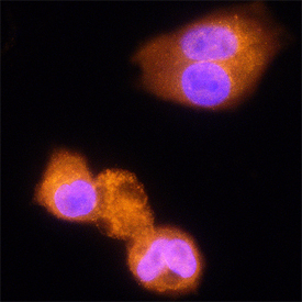
- Main image

- Experimental details
- alpha-L-Iduronidase/IDUA in HepG2 Human Cell Line. alpha-L-Iduronidase/IDUA was detected in immersion fixed HepG2 human hepatocellular carcinoma cell line using Sheep Anti-Human alpha-L-Iduronidase/IDUA Antigen Affinity-purified Polyclonal Antibody (Catalog # AF4119) at 15 µg/mL for 3 hours at room temperature. Cells were stained using the NorthernLights™ 557-conjugated Anti-Sheep IgG Secondary Antibody (red; Catalog # NL010) and counterstained with DAPI (blue). Specific staining was localized to cytoplasm. View our protocol for Fluorescent ICC Staining of Cells on Coverslips.