Explore
 Validate
 Learn
Learn Western blot
 ELISA
ELISAAntibody data
- Antibody Data
- Antigen structure
- References [0]
- Comments [0]
- Validations
- Western blot [1]
- Immunocytochemistry [2]
- Immunohistochemistry [1]
Submit
Validation data
Reference
Comment
Report error
- Product number
- 200-301-390 - Provider product page

- Provider
- Invitrogen Antibodies
- Product name
- Keratin Monoclonal Antibody (C11)
- Antibody type
- Monoclonal
- Antigen
- Purifed from natural sources
- Reactivity
- Human
- Host
- Mouse
- Isotype
- IgG
- Antibody clone number
- C11
- Vial size
- 100 µg
- Concentration
- 1.3 mg/mL
- Storage
- -20° C, Avoid Freeze/Thaw Cycles
No comments: Submit comment
Supportive validation
- Submitted by
- Invitrogen Antibodies (provider)
- Main image

- Experimental details
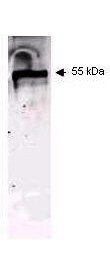
- Western blot using ROCKLAND Immunochemicals Mouse Anti-Keratin antibody. This antibody recognizes a single 56 kDa band corresponding to human keratin as confirmed by the position of molecular weight markers (not shown). Approximately 100 ng of keratin from human epidermis (Sigma p/n K0253) was applied under reducing conditions to a pre-cast 4-20% iGel from Gradipore Inc. A 1:400 dilution of Mab anti-Keratin was used for 2h followed by detection using a 1:5,000 dilution of IRDyeTM800 conjugated Goat-a-Mouse IgG [H&L] (610-132-121) and visualization using the Odyssey® Infrared Imaging System developed by LI-COR. Other detection systems will yield similar results. IRDye is a trademark of LI-COR, Inc.
Supportive validation
- Submitted by
- Invitrogen Antibodies (provider)
- Main image
- Experimental details
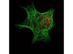
- Immunofluorescence Microscopy of Rockland Immunochemicals Anti-Keratin antibody (200-301-390) was used with Rocklands Dylight 488 goat anti-mouse 610-141-121 (shown in green) to detect Keratin by Immunofluorescence. In the same experiment, Rocklands polyclonal Anti-HDAC-1 antibody (600-401-879) was used with Atto425 Anti-Rabbit IgG 611-151-122 (shown in red) to detect HDAC-1. Data was collected on a STED-CW TCS-SP5 Confocal system (Leica Microsystems) equipped with a DFC 350FX camera allowing sequential acquisition in wide-field, confocal and STED CW imaging modes and provided courtesy of: Myriam Gastard, PhD, personal communication, Leica Microsystems, Inc. USA

- Submitted by
- Invitrogen Antibodies (provider)
- Main image
- Experimental details
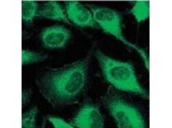
- Immunofluorescence using ROCKLAND Immunochemicals Mouse Anti-Keratin antibody. Confocal slices of HeLa cells are between 0.5 and 0.6 µm where the image is taken near the bottom of the cell. Use FITC a 1:2,000 dilution of FITC conjugated Goat-a-Mouse IgG [H&L] (610-102-121) for detection

Supportive validation
- Submitted by
- Invitrogen Antibodies (provider)
- Main image
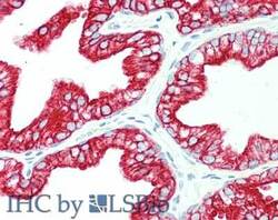
- Experimental details
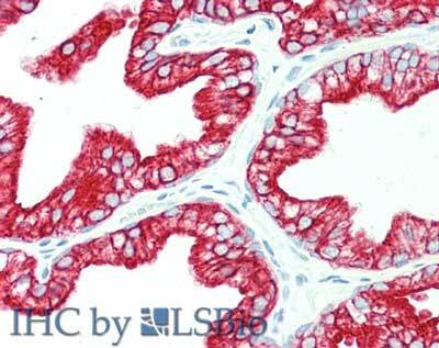
- Immunohistochemistry of Mouse anti-Keratin antibody. Tissue: human prostate. Fixation: formalin fixed paraffin embedded. Antigen retrieval: not required. Primary antibody: anti-Keratin antibody at 10 µg/mL for 1 h at RT. Secondary antibody: Peroxidase mouse secondary antibody at 1:10,000 for 45 min at RT. Staining: Keratin as precipitated red signal with hematoxylin purple nuclear counterstain.