 Explore
 Validate
 Learn
Learn730019
antibody from Invitrogen Antibodies
Targeting: CBX5
HP1, HP1-ALPHA, HP1Hs-alpha
 Western blot
 Immunocytochemistry Immunoprecipitation Immunohistochemistry Flow cytometry Chromatin Immunoprecipitation Other assay
Antibody data
- Antibody Data
- Antigen structure
- References [1]
- Comments [0]
- Validations
- Immunocytochemistry [2]
- Immunohistochemistry [1]
- Flow cytometry [1]
- Chromatin Immunoprecipitation [1]
- Other assay [2]
Submit
Validation data
Reference
Comment
Report error
- Product number
- 730019 - Provider product page

- Provider
- Invitrogen Antibodies
- Product name
- Anti-HP1 alpha Monoclonal Antibody
- Antibody type
- Monoclonal
- Antigen
- Other
- Reactivity
- Human, Mouse, Rat
- Host
- Mouse
- Isotype
- IgG
- Vial size
- 100 µg
- Concentration
- 0.5 mg/ml
- Storage
- Maintain refrigerated at 2-8°C for up to 1 month. For long term storage store at -20°C
Submitted references Assessment of a method to characterize antibody selectivity and specificity for use in immunoprecipitation.
Marcon E, Jain H, Bhattacharya A, Guo H, Phanse S, Pu S, Byram G, Collins BC, Dowdell E, Fenner M, Guo X, Hutchinson A, Kennedy JJ, Krastins B, Larsen B, Lin ZY, Lopez MF, Loppnau P, Miersch S, Nguyen T, Olsen JB, Paduch M, Ravichandran M, Seitova A, Vadali G, Vogelsang MS, Whiteaker JR, Zhong G, Zhong N, Zhao L, Aebersold R, Arrowsmith CH, Emili A, Frappier L, Gingras AC, Gstaiger M, Paulovich AG, Koide S, Kossiakoff AA, Sidhu SS, Wodak SJ, Gräslund S, Greenblatt JF, Edwards AM
Nature methods 2015 Aug;12(8):725-31
Nature methods 2015 Aug;12(8):725-31
No comments: Submit comment
Supportive validation
- Submitted by
- Invitrogen Antibodies (provider)
- Main image
- Experimental details
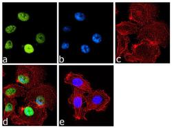
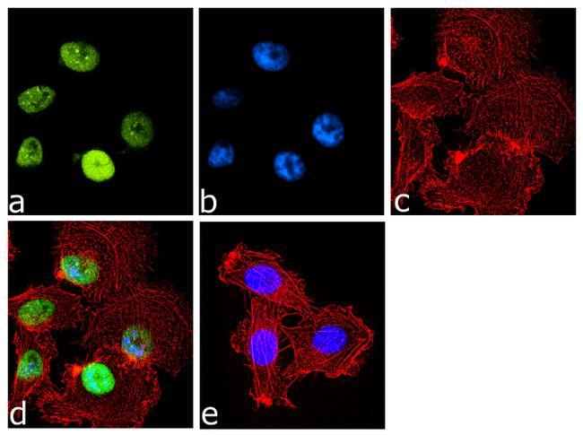
- Immunofluorescence analysis of CBX5 was done on 70% confluent log phase PC-3 cells. The cells were fixed with 4% paraformaldehyde for 10 minutes, permeabilized with 0.1% Triton™ X-100 for 10 minutes, and blocked with 1% BSA for 1 hour at room temperature. The cells were labeled with CBX5 Mouse Monoclonal Antibody (Product # 730019) at 2 µg/mL in 0.1% BSA and incubated for 3 hours at room temperature and then labeled with Goat anti-Mouse IgG (H+L) Superclonal™ Secondary Antibody, Alexa Fluor® 488 conjugate (Product # A28175) at a dilution of 1:2000 for 45 minutes at room temperature (Panel a: green). Nuclei (Panel b: blue) were stained with SlowFade® Gold Antifade Mountant with DAPI (Product # S36938). F-actin (Panel c: red) was stained with Alexa Fluor® 555 Rhodamine Phalloidin (Product # R415, 1:300). Panel d is a merged image showing nuclear localization. Panel e is a no primary antibody control. The images were captured at 60X magnification.
- Submitted by
- Invitrogen Antibodies (provider)
- Main image
- Experimental details
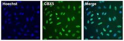
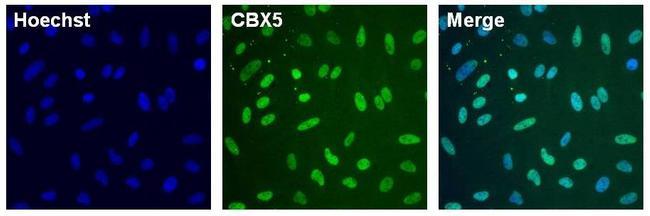
- Immunofluorescent analysis of CBX5 (green) in HeLa cells. The cells were fixed with formalin for 15 minutes, permeabilized with 1X Permeablilization buffer (Product # 8408400) for 15 minutes, and blocked with 1% Blocker BSA (Product # 37525) for 15 minutes at room temperature. Cells were stained with CBX5 monoclonal antibody (Product # 730019) at a dilution of 1:250 in blocking buffer for one hour at room temperature, washed with 1X TBS Tween 20 Buffer (Product # 28360), followed by incubation with DyLight 488 goat anti-mouse IgG secondary antibody (green, Product # A28175) at a dilution of 1:2000 and Hoechst 33342 dye (blue, Product # 62249) at a dilution of 1:5000 for 30 minutes at room temperature. Images were taken on a Thermo Scientific ToxInsight at 20X magnification.
Supportive validation
- Submitted by
- Invitrogen Antibodies (provider)
- Main image
- Experimental details
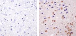
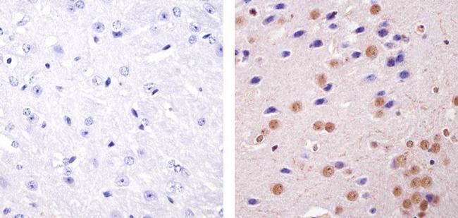
- Immunohistochemistry analysis of CBX5 showing staining in the nucleus of paraffin-embedded mouse brain tissue (right) compared to a negative control without primary antibody (left). To expose target proteins, antigen retrieval was performed using 10mM sodium citrate (pH 6.0), microwaved for 8-15 min. Following antigen retrieval, tissues were blocked in 3% H2O2-methanol for 15 min at room temperature, washed with ddH2O and PBS, and then probed with a Anti- CBX5 Monoclonal Antibody (Product # 730019) diluted in 3% BSA-PBS at a dilution of 1:100 overnight at 4°C in a humidified chamber. Tissues were washed extensively in PBST and detection was performed using an HRP-conjugated secondary antibody followed by colorimetric detection using a DAB kit. Tissues were counterstained with hematoxylin and dehydrated with ethanol and xylene to prep for mounting.
Supportive validation
- Submitted by
- Invitrogen Antibodies (provider)
- Main image
- Experimental details
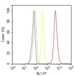
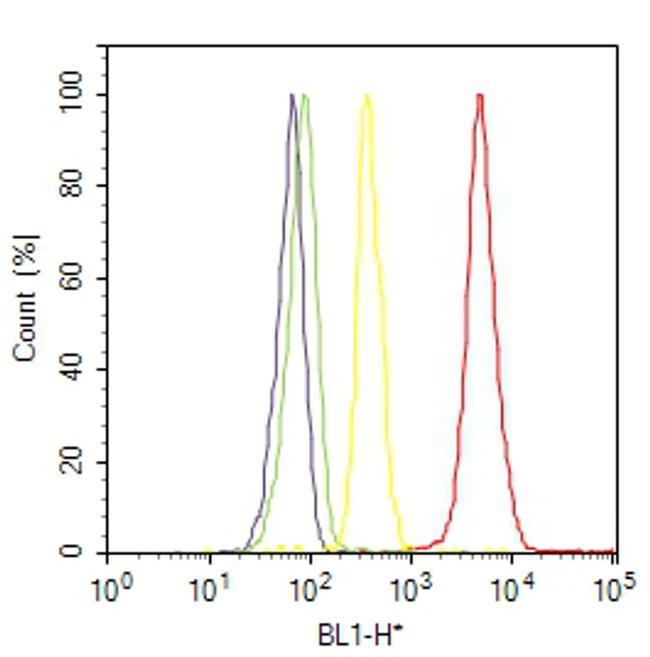
- Flow cytometry analysis of CBX5 was done on HT-29 cells. Cells were fixed with 70% ethanol for 10 minutes, permeabilized with 0.25% Triton™ X-100 for 20 minutes, and blocked with 5% BSA for 30 minutes at room temperature. Cells were labeled with CBX5 Mouse Monoclonal Antibody (730019, red histogram) or with mouse isotype control (yellow histogram) at 3-5 ug/million cells in 2.5% BSA. After incubation at room temperature for 2 hours, the cells were labeled with Alexa Fluor® 488 Rabbit Anti-Mouse Secondary Antibody (A11059) at a dilution of 1:400 for 30 minutes at room temperature. The representative 10,000 cells were acquired and analyzed for each sample using an Attune® Acoustic Focusing Cytometer. The purple histogram represents unstained control cells and the green histogram represents no-primary-antibody control.
Supportive validation
- Submitted by
- Invitrogen Antibodies (provider)
- Main image
- Experimental details
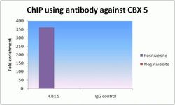
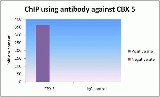
- ChIP qPCR analysis of CBX 5 Monoclonal Antibody, Recombinant (Product # 730019). ChIP was performed on HeLa chromatin using CBX 5 Mouse Monoclonal Antibody. Realtime PCR was performed for MYT and negative control locus targets using Isotype antibody as a negative control. Data is presented as fold enrichment of the ChIP antibody signal versus the negative control IgG using the comparative CT method.
Supportive validation
- Submitted by
- Invitrogen Antibodies (provider)
- Main image
- Experimental details
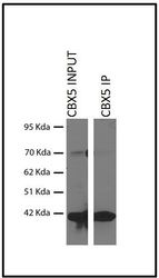
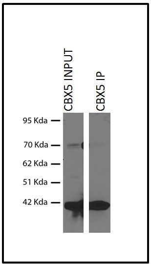
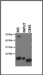
- Immunoprecipitation analysis of Anti-CBX5 Recombinant Mouse Monoclonal Antibody (Product # 730019). Immunoprecipitation analysis was performed on HEK293 cells over expressing flag tagged CBX5 protein. Western blot using anti-flag antibody was performed on the raw lysate.
- Submitted by
- Invitrogen Antibodies (provider)
- Main image
- Experimental details
- Immunoprecipitation analysis of Anti-CBX5 Recombinant Mouse Monoclonal Antibody (Product # 730019). Immunoprecipitation analysis was performed on HEK293 cells over expressing flag tagged CBX5 protein. Western blot using anti-flag antibody was performed on the raw lysate.
