Explore
 Validate
 Learn
Learn Western blot
Antibody data
- Antibody Data
- Antigen structure
- References [0]
- Comments [0]
- Validations
- Western blot [3]
- Immunocytochemistry [1]
Submit
Validation data
Reference
Comment
Report error
- Product number
- PA1-16945 - Provider product page

- Provider
- Invitrogen Antibodies
- Product name
- PDK1 Polyclonal Antibody
- Antibody type
- Polyclonal
- Antigen
- Synthetic peptide
- Description
- Suggested positive control: human heart protein, antigen standard for PDK1 (transient overexpression lysate).
- Reactivity
- Human, Mouse
- Host
- Rabbit
- Isotype
- IgG
- Vial size
- 100 µL
- Concentration
- 0.5 mg/mL
- Storage
- -20° C, Avoid Freeze/Thaw Cycles
No comments: Submit comment
Supportive validation
- Submitted by
- Invitrogen Antibodies (provider)
- Main image

- Experimental details
- Detection of PDK1 in human heart lysate using Product # PA1-16945 at 0.5 µg/mL.

- Submitted by
- Invitrogen Antibodies (provider)
- Main image

- Experimental details
- Western blot analysis of PDK1 in human heart lysate. Samples were incubated in PDK1 polyclonal antibody (Product # PA1-16945) using a dilution of 0.5 µg/mL.

- Submitted by
- Invitrogen Antibodies (provider)
- Main image

- Experimental details
- Western blot was performed using Anti-Pyruvate dehydrogenase kinase isozyme 1 protein Polyclonal Antibody (Product # PA1-16945) and 49.4 kDa band corresponding to Pyruvate dehydrogenase kinase isozyme 1 protein was observed. Hypoxia conditions such as cobalt chloride and deferoxamine mesylate treatments are known to increase the expression of Pyruvate dehydrogenase kinase isozyme 1 protein. Whole cell extracts (30 µg lysate) of HeLa (Lane 1), HeLa treated with cobalt chloride (150uM for 24 hr) (Lane 2), HeLa treated with deferoxamine mesylate (10uM for 20 hr) (Lane 3), Mouse heart (Lane 4) and Mouse liver (Lane 5) and Rat heart (Lane 6) were electrophoresed using Novex® NuPAGE® 4-12% Bis-Tris gel (Product # NP0342BOX). Resolved proteins were then transferred onto a nitrocellulose membrane (Product # IB23001) by iBlot® 2 Dry Blotting System (Product # IB21001). The blot was probed with the primary antibody (1:1000 dilution) and detected by Goat anti-Rabbit IgG (H+L) Superclonal™ Recombinant Secondary Antibody, HRP conjugate (Product # A27036, 0.25 µg/ml, 1:4000 dilution) using the iBright FL 1000 (Product # A32752). Chemiluminescent detection was performed using Novex® ECL Chemiluminescent Substrate Reagent Kit (Product # WP20005).

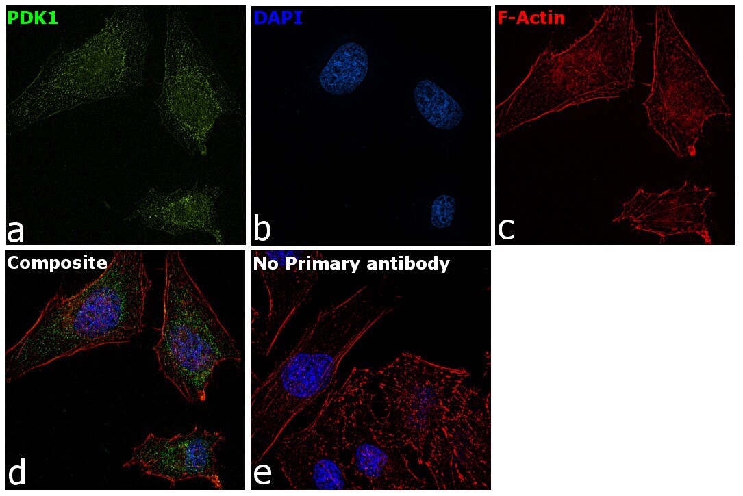
Supportive validation
- Submitted by
- Invitrogen Antibodies (provider)
- Main image
- Experimental details
- Immunofluorescence analysis of PDK1 was performed using 70% confluent log phase HeLa cells. The cells were fixed with 4% paraformaldehyde for 10 minutes, permeabilized with 0.1% Triton™ X-100 for 15 minutes, and blocked with 2% BSA for 1 hour at room temperature. The cells were labeled with PDK1 Polyclonal Antibody (Product # PA1-16945) at 5 µg/mL in 0.1% BSA, incubated at 4 degree Celsius overnight and then labeled with Goat anti-Rabbit IgG (H+L) Superclonal™ Secondary Antibody, Alexa Fluor® 488 conjugate (Product # A27034) at a dilution of 1:2000 for 45 minutes at room temperature (Panel a: green). Nuclei (Panel b: blue) were stained with ProLong™ Diamond Antifade Mountant with DAPI (Product # P36962). F-actin (Panel c: red) was stained with Rhodamine Phalloidin (Product # R415). Panel d represents the merged image showing Cytoplasmic localization. Panel e represents control cells with no primary antibody to assess background. The images were captured at 60X magnification.