Explore
 Validate
 Learn
Learn Western blot
Antibody data
- Antibody Data
- Antigen structure
- References [1]
- Comments [0]
- Validations
- Western blot [2]
- Immunohistochemistry [3]
Submit
Validation data
Reference
Comment
Report error
- Product number
- MAB3320 - Provider product page

- Provider
- Novus Biologicals
- Product name
- Mouse Monoclonal Calbindin D/S100G Antibody
- Antibody type
- Monoclonal
- Description
- Protein A or G purified from hybridoma culture supernatant. Detects human Calbindin D in direct ELISAs and Western blots. In direct ELISAs and Western blots, this antibody does not cross-react with recombinant human S100P.
- Reactivity
- Human
- Host
- Mouse
- Conjugate
- Unconjugated
- Isotype
- IgG
- Vial size
- 100 ug
- Concentration
- LYOPH
- Storage
- Use a manual defrost freezer and avoid repeated freeze-thaw cycles. 12 months from date of receipt, -20 to -70 degreesC as supplied. 1 month, 2 to 8 degreesC under sterile conditions after reconstitution. 6 months, -20 to -70 degreesC under sterile conditions after reconstitution.
Submitted references Cerebrospinal Fluid Calbindin D Concentration as a Biomarker of Cerebellar Disease Progression in Niemann-Pick Type C1 Disease.
Bradbury A, Bagel J, Sampson M, Farhat N, Ding W, Swain G, Prociuk M, O'Donnell P, Drobatz K, Gurda B, Wassif C, Remaley A, Porter F, Vite C
The Journal of pharmacology and experimental therapeutics 2016 Aug;358(2):254-61
The Journal of pharmacology and experimental therapeutics 2016 Aug;358(2):254-61
No comments: Submit comment
Supportive validation
- Submitted by
- Novus Biologicals (provider)
- Main image
- Experimental details
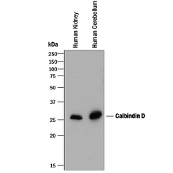
- Detection of Human Calbindin D by Western Blot. Western blot shows lysates of human kidney tissue and human brain (cerebellum) tissue. PVDF membrane was probed with 0.25 µg/mL of Mouse Anti-Human Calbindin D Monoclonal Antibody (Catalog # MAB3320) followed by HRP-conjugated Anti-Mouse IgG Secondary Antibody (Catalog # HAF018). A specific band was detected for Calbindin D at approximately 28 kDa (as indicated). This experiment was conducted under reducing conditions and using Immunoblot Buffer Group 1.

- Submitted by
- Novus Biologicals (provider)
- Main image

- Experimental details
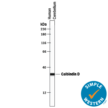
- Detection of Human Calbindin D by Simple WesternTM. Simple Western lane view shows lysates of human brain (cerebellum) tissue, loaded at 0.2 mg/mL. A specific band was detected for Calbindin D at approximately 32 kDa (as indicated) using 10 µg/mL of Mouse Anti-Human Calbindin D Monoclonal Antibody (Catalog # MAB3320) . This experiment was conducted under reducing conditions and using the 12-230 kDa separation system.
Supportive validation
- Submitted by
- Novus Biologicals (provider)
- Main image
- Experimental details
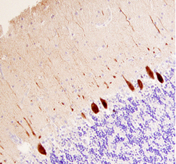
- Calbindin D in Rat Brainstem. Calbindin D in Rat Brainstem.
- Submitted by
- Novus Biologicals (provider)
- Main image
- Experimental details
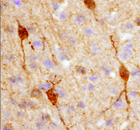
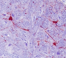
- Calbindin D in Human Brain. Calbindin D was detected in immersion fixed paraffin-embedded sections of human brain (cerebellum) using 25 µg/mL Mouse Anti-Human Calbindin D Monoclonal Antibody (Catalog # MAB3320) overnight at 4 °C. Tissue was stained with the Anti-Mouse HRP-DAB Cell & Tissue Staining Kit (brown; Catalog # CTS002) and counterstained with hematoxylin (blue). View our protocol for Chromogenic IHC Staining of Paraffin-embedded Tissue Sections.

- Submitted by
- Novus Biologicals (provider)
- Main image
- Experimental details
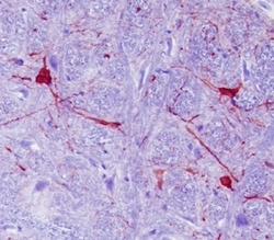
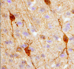
- Calbindin D in Rat Brain. Calbindin D was detected in perfusion fixed frozen sections of rat brain (hypothalamus) using 25 µg/mL Mouse Anti-Human Calbindin D Monoclonal Antibody (Catalog # MAB3320) overnight at 4 °C. Tissue was stained with the Anti-Mouse HRP-DAB Cell & Tissue Staining Kit (brown; Catalog # CTS002) and counterstained with hematoxylin (blue). View our protocol for Chromogenic IHC Staining of Frozen Tissue Sections.