 Explore
 Validate
 Learn
Learn Western blot
 Immunohistochemistry
Antibody data
- Antibody Data
- Antigen structure
- References [1]
- Comments [0]
- Validations
- Immunohistochemistry [1]
- Flow cytometry [2]
- Other assay [1]
Submit
Validation data
Reference
Comment
Report error
- Product number
- PA5-26383 - Provider product page

- Provider
- Invitrogen Antibodies
- Product name
- HSL Polyclonal Antibody
- Antibody type
- Polyclonal
- Antigen
- Synthetic peptide
- Reactivity
- Human
- Host
- Rabbit
- Isotype
- IgG
- Vial size
- 400 μL
- Concentration
- 2 mg/mL
- Storage
- Store at 4°C short term. For long term storage, store at -20°C, avoiding freeze/thaw cycles.
Submitted references Secretion of autoimmune antibodies in the human subcutaneous adipose tissue.
Frasca D, Diaz A, Romero M, Thaller S, Blomberg BB
PloS one 2018;13(5):e0197472
PloS one 2018;13(5):e0197472
No comments: Submit comment
Supportive validation
- Submitted by
- Invitrogen Antibodies (provider)
- Main image

- Experimental details
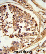
- Immunohistochemistry analysis of HSL in formalin-fixed and paraffin-embedded human testis tissue. Samples were incubated with HSL polyclonal antibody (Product # PA5-26383) which was peroxidase-conjugated to the secondary antibody, followed by DAB staining. This data demonstrates the use of this antibody for immunohistochemistry; clinical relevance has not been evaluated.
Supportive validation
- Submitted by
- Invitrogen Antibodies (provider)
- Main image

- Experimental details
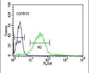
- Flow cytometry analysis of HL-60 cells using a LIPE polyclonal antibody (Product # PA5-26383) (right) compared to a negative control cell (left) at a dilution of 1:10-50, followed by a FITC-conjugated goat anti-rabbit antibody
- Submitted by
- Invitrogen Antibodies (provider)
- Main image

- Experimental details
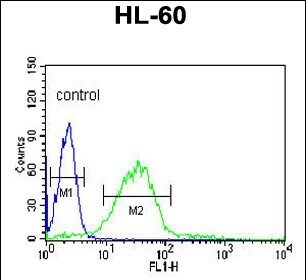
- Flow cytometry of HSL in HL-60 cells (right histogram). Samples were incubated with HSL polyclonal antibody (Product # PA5-26383) followed by FITC-conjugated goat-anti-rabbit secondary antibody. Negative control cell (left histogram).
Supportive validation
- Submitted by
- Invitrogen Antibodies (provider)
- Main image
- Experimental details
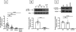
- Fig 5 Measure of lipolysis in the obese SAT versus blood. A. Adipocytes (AD), SVF and PBMC (blood) were sonicated for cell disruption in the presence of TRIzol to separate the soluble fraction (used for RNA isolation) from lipids and cell debris. AD and SVF were from the same obese individuals. PBMC (blood) were from obese individuals age-, gender- and BMI-matched. Results show qPCR values (2 -DeltaDeltaCt ) of LPL RNA expression. B. Total protein lysates of adipocytes (AD) and SVF (from different obese individuals age-, gender- and BMI-matched) were prepared and run in WB to measure phospho-HSL. A representative WB for the higest and lowest values is shown (top). Mean comparisons between groups were performed by Student's t test (two-tailed). ***p
